

Anaplasma phagocytophilum in Marmota himalayana
- PMID: 35490230
- PMCID: PMC9055747
- DOI: 10.1186/s12864-022-08557-x
Anaplasma phagocytophilum in Marmota himalayana
Abstract
Background: Human granulocytic anaplasmosis is a tick-borne zoonotic disease caused by Anaplasma phagocytophilum. Coinfections with A. phagocytophilum and other tick-borne pathogens are reported frequently, whereas the relationship between A. phagocytophilum and flea-borne Yersnia pestis is rarely concerned.
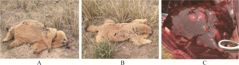
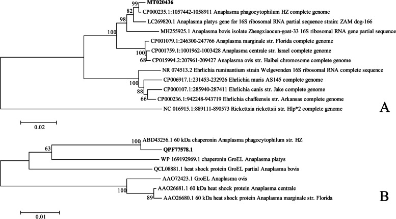
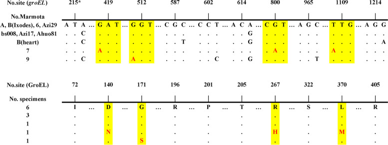
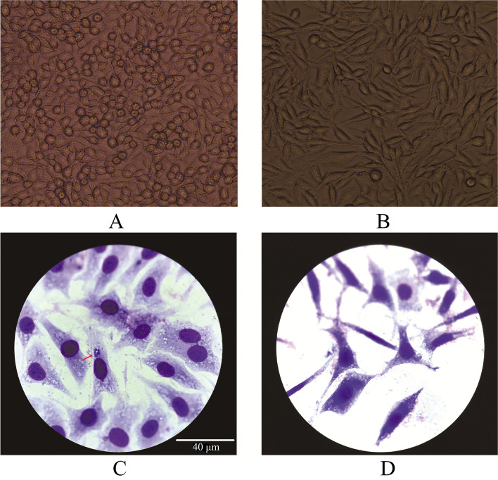
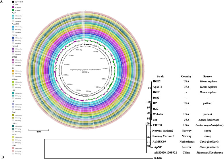
Results: A. phagocytophilum and Yersnia pestis were discovered within a Marmota himalayana found dead in the environment, as determined by 16S ribosomal rRNA sequencing. Comparative genomic analyses of marmot-derived A. phagocytophilum isolate demonstrated its similarities and a geographic isolation from other global strains. The 16S rRNA gene and GroEL amino acid sequence identity rates between marmot-derived A. phagocytophilum (JAHLEX000000000) and reference strain HZ (CP000235.1) are 99.73% (1490/1494) and 99.82% (549/550), respectively. 16S rRNA and groESL gene screenings show that A. phagocytophilum is widely distributed in marmots; the bacterium was more common in marmots found dead (24.59%, 15/61) than in captured marmots (19.21%, 29/151). We found a higher Y. pestis isolation rate in dead marmots harboring A. phagocytophilum than in those without it (2 = 4.047, p < 0.05). Marmot-derived A. phagocytophilum was able to live in L929 cells and BALB/c mice but did not propagate well.
Conclusions: In this study, A. phagocytophilum was identified for the first time in Marmota himalayana, a predominant Yersinia pestis host. Our results provide initial evidence for M. himalayana being a reservoir for A. phagocytophilum; moreover, we found with the presence of A. phagocytophilum, marmots may be more vulnerable to plague. Humans are at risk for co-infection with both pathogens by exposure to such marmots.
Keywords: Anaplasma phagocytophilum; Anaplasmosis; Coinfection; Marmota himalayana; Plague; Yersinia pestis.
© 2022. The Author(s).
Conflict of interest statement
The authors declare that they have no competing interests.
Figures
References
-
- Liu Y, Tian J, Shen E. The atlas of plague and its environment in People’s Republic of China. Beijing: Science Press; 2000.
MeSH terms
Substances
Grants and funding
LinkOut - more resources
Full Text Sources
Research Materials
