. 2022 Aug 3;30(8):2680-2692.
doi: 10.1016/j.ymthe.2022.04.020. Epub 2022 Apr 30.

Paired nicking-mediated COL17A1 reframing for junctional epidermolysis bullosa

Affiliations

Paired nicking-mediated COL17A1 reframing for junctional epidermolysis bullosa

Johannes Bischof et al. Mol Ther. .

Abstract

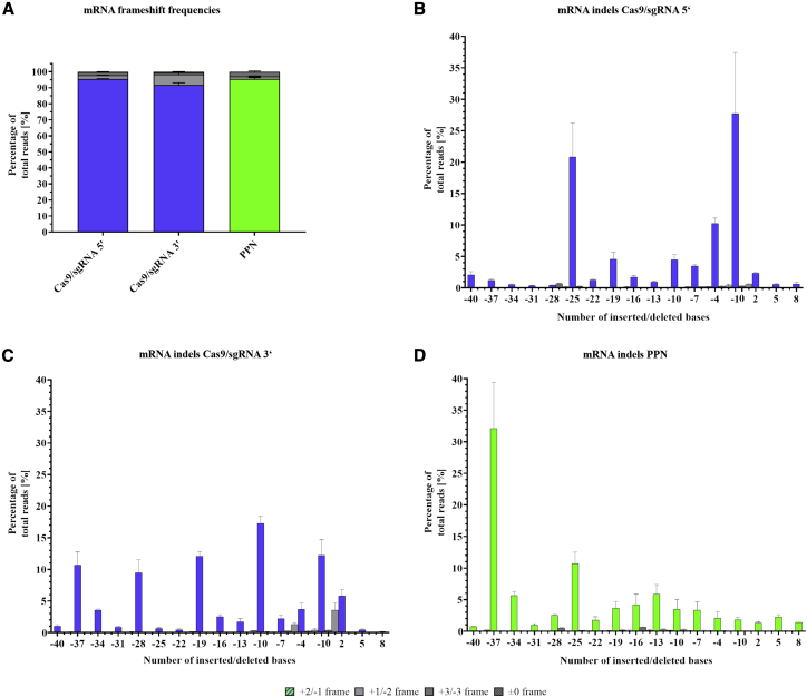
Junctional epidermolysis bullosa (JEB) is a debilitating hereditary skin disorder caused by mutations in genes encoding laminin-332, type XVII collagen (C17), and integrin-α6β4, which maintain stability between the dermis and epidermis. We designed patient-specific Cas9-nuclease- and -nickase-based targeting strategies for reframing a common homozygous deletion in exon 52 of COL17A1 associated with a lack of full-length C17 expression. Subsequent characterization of protein restoration, indel composition, and divergence of DNA and mRNA outcomes after treatment revealed auspicious efficiency, safety, and precision profiles for paired nicking-based COL17A1 editing. Almost 46% of treated primary JEB keratinocytes expressed reframed C17. Reframed COL17A1 transcripts predominantly featured 25- and 37-nt deletions, accounting for >42% of all edits and encoding C17 protein variants that localized accurately to the cell membrane. Furthermore, corrected cells showed accurate shedding of the extracellular 120-kDa C17 domain and improved adhesion capabilities to laminin-332 compared with untreated JEB cells. Three-dimensional (3D) skin equivalents demonstrated accurate and continuous deposition of C17 within the basal membrane zone between epidermis and dermis. Our findings constitute, for the first time, gene-editing-based correction of a COL17A1 mutation and demonstrate the superiority of proximal paired nicking strategies based on Cas9 D10A nickase over wild-type Cas9-based strategies for gene reframing in a clinical context.

Keywords: CRISPR-Cas9; JEB; gene reframing; junctional epidermolysis bullosa; next-generation sequencing; paired nicking; skin equivalents.


Conflict of interest statement

Declaration of interests T.C. and S.A.H. have filed a patent application for Abnoba-Seq. T.C. has a sponsored research collaboration with Cellectis and is an advisor to Cimeo Therapeutics and Excision BioTherapeutics. The other authors declare no competing interests.

Figures

Figure 1
Figure 1
Gene-reframing strategy for COL17A1 repair via PPN-mediated DNA cleavage sgRNAs were designed to specifically target exon 52 of COL17A1. The target sites of sgRNAs 3′ and 5′ overlap by 1 bp (highlighted in blue). While sgRNA 5′ is not mutation-specific, the target site of sgRNA 3′ covers the patient mutation (c.3899_3900delCT). As targeting of the wild-type sequence requires tolerance of a 2-bp DNA bulge, this sgRNA is expected to demonstrate a high degree of allelic specificity. Since the sgRNA 3′ Cas9 cleavage site is adjacent to the mutation (ΔCT), this will generate reframing indels with the least impact on the aa code. Created with BioRender.com.
Figure 2
Figure 2
DNA frameshift frequencies and indel patterns from Cas9/sgRNA 5′-, Cas9/sgRNA 3′-, and PPN-treated primary JEB keratinocytes (A) Frequencies of the three possible reading frames of COL17A1 observed in DNA-derived amplicons after treatment of JEB keratinocytes. (B–D) Analysis of indel patterns after treatment of primary JEB keratinocytes with (B) Cas9/sgRNA 5′, (C) Cas9/sgRNA 3′, or (D) PPN. Indels ranging from -40 to +8 bp have been plotted, accounting for the vast majority of all indels generated. (A–D) Restored COL17A1 reading frames (+2/-1) are indicated as blue (wild-type Cas9) or green (Cas9 nickase) for each sample. Reading frames analogous to patient keratinocytes (+3/-3) are colored dark gray, and alternative reading frames (+1/-2) are colored light gray. The gray hashed bars constitute a subset of the +3/-3 reading frames. These alleles are either untargeted or subject to perfect DSB repair (±0). n = 3; mean ± SEM; for statistical analysis of (A), please refer to Table S1A.
Figure 3
Figure 3
mRNA frameshift frequencies and indel patterns from Cas9/sgRNA 5′-, Cas9/sgRNA 3′-, and PPN-treated primary JEB keratinocytes (A) Frequencies of the three possible reading frames of COL17A1 observed in mRNA-derived amplicons after treatment of JEB keratinocytes. (B–D) Analysis of indel patterns after treatment of primary JEB keratinocytes with (B) Cas9/sgRNA 5′, (C) Cas9/sgRNA 3′, or (D) PPN. Indels ranging from -40 to +8 nucleotides have been plotted, accounting for the vast majority of all indels generated. (A–D) Restored COL17A1 reading frames (+2/-1) are indicated as blue (wild-type Cas9) or green (Cas9 nickase) for each sample. Reading frames analogous to patient keratinocytes (+3/-3) are colored dark gray, and alternative reading frames (+1/-2) are colored light gray. The gray hashed bars constitute a subset of the +3/-3 reading frames. These alleles are either untargeted or subject to perfect DSB repair (±0). n = 3; mean ± SEM; for statistical analysis of (A), please refer to Table S1B.
Figure 4
Figure 4
NGS-based off-target analysis for sgRNA 5′ and 3′ (A) Heatmap and observed frequencies of indel formation at predicted sgRNA 5′ off targets. Off-target regions were amplified from untreated (control) as well as Cas9/sgRNA 5′- and PPN-treated JEB cells. (B) Heatmap and observed frequencies of indels at predicted sgRNA 3′ off targets. Off-target regions were amplified from untreated (control) as well as Cas9/sgRNA 3′- and PPN-treated JEB cells.
Figure 5
Figure 5
Phenotypic analysis of corrected, primary JEB keratinocytes (A) Flow cytometric analysis revealed high C17 positivity in single Cas9- and PPN-treated cells. Primary wild-type and untreated JEB cells served as controls. n = 3; mean ± SEM; one-way ANOVA with Dunnett’s multiple comparisons test. (B) sqRT-PCR of treated keratinocytes indicated strong transcript rescue from NMD in all treatments. Wild-type and untreated JEB cells served as controls. n = 3; mean ± SEM; one-way ANOVA with Dunnett’s multiple comparisons test. (C) Western blot analysis of cell lysates showed highly efficient restoration of C17 (180 kDa) in PPN-treated primary cells. C17 levels of corrected cells were similar to those of wild-type cells. Bands were normalized to wild-type keratinocytes and β-tubulin (50 kDa). (D) Western blot (WB) analysis of cell supernatants confirmed the correct shedding of the extracellular C17 domain (120 kDa) in all treatments. Bands were normalized to wild-type keratinocytes. Mean percentages from densitometric WB analysis are given in the table below the blots. n = 3. (E) Representative immunofluorescence stainings for C17 (green) in combination with DAPI (blue). Staining was conducted without the use of permeabilizing reagents, confirming the correct localization of C17 in the cell membrane. Images show 2 × 2 tile scans of the 20× objective as well as single scans of the 40× objective.
Figure 6
Figure 6
Laminin-332 adhesion assay of PPN-corrected JEB keratinocytes (A) Indel outcome and allele diversity for the single-cell clones used for the adhesion assay of immortalized, PPN-treated JEB keratinocytes. aa similar to the wild-type sequence are displayed in black, and those similar to the patient/alternative reading frame are displayed in red. Deletions are indicated by spaces, and stop codons are indicated at the end of the sequence. (B) The adhesive strength of several cell lines and clones was tested with laminin-332-precoated tissue culture plates. Bulk as well as single-cell clones of PPN-treated immortalized JEB keratinocytes showed higher adhesion when compared with untreated cells. n = 3; mean ± SEM; one-way ANOVA with Dunnett’s multiple comparisons test. (C) Analysis of primary keratinocytes revealed a generally stronger adhesive strength when compared with immortalized keratinocytes. The amount of PPN-treated primary JEB keratinocytes still attached to the coated surface after mechanical stress is significantly higher when compared with untreated patient keratinocytes. n = 3; mean ± SEM; unpaired Student’s t test. For extended statistical analysis of (B) and (C), please refer to Tables S1C and S1D, respectively.
Figure 7
Figure 7
C17 and laminin-332 IF stainings of 3D skin equivalents from immortalized JEB keratinocytes with PPN treatment Immunofluorescence staining for C17 (green) in combination with laminin-332 (red) and DAPI (blue). Epidermis and dermis are marked with “E” and “D”, respectively. (A and B) Skin equivalents of (A) positive (wild-type) and (B) negative control (JEB) cells. (C and D) Skin equivalents constructed from PPN-treated JEB keratinocytes, unsorted (C) and sorted (D). Images show 1 × 3 tile scans of the 10× objective as well as single scans of the 40× objective.

References

    1. March O.P., Kocher T., Koller U. Context-dependent strategies for enhanced genome editing of genodermatoses. Cells. 2020;9:112. doi: 10.3390/cells9010112. - DOI - PMC - PubMed
    1. March O.P., Reichelt J., Koller U. Gene editing for skin diseases: designer nucleases as tools for gene therapy of skin fragility disorders. Exp. Physiol. 2018;103:449–455. doi: 10.1113/EP086044. - DOI - PubMed
    1. Kocher T., Koller U. Advances in gene editing strategies for epidermolysis bullosa. Prog. Mol. Biol. Transl. Sci. 2021;182:81–109. doi: 10.1016/bs.pmbts.2020.12.007. - DOI - PubMed
    1. Kocher T., Petkovic I., Bischof J., Koller U. Current developments in gene therapy for epidermolysis bullosa. Expert Opin. Biol. Ther. 2022:1–14. doi: 10.1080/14712598.2022.2049229. - DOI - PubMed
    1. Sfeir A., Symington L.S. Microhomology-mediated end joining: a back-up survival mechanism or dedicated pathway? Trends Biochem. Sci. 2015;40:701–714. doi: 10.1016/j.tibs.2015.08.006. - DOI - PMC - PubMed

Publication types