

Eosinophilic Fasciitis with Hypereosinophilia as the Initial Clinical Manifestation of Peripheral T-Cell Lymphoma, Not Otherwise Specified
- PMID: 35491131
- PMCID: PMC9751711
- DOI: 10.2169/internalmedicine.9300-21
Eosinophilic Fasciitis with Hypereosinophilia as the Initial Clinical Manifestation of Peripheral T-Cell Lymphoma, Not Otherwise Specified
Abstract
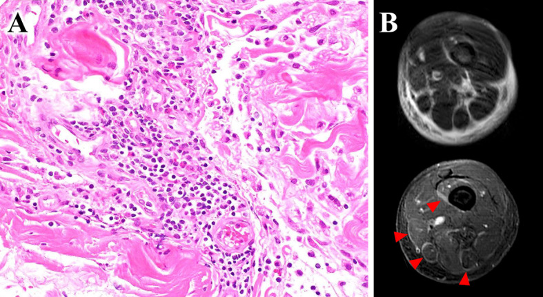
A 58-year-old man presented with painful edema of the extremities, and a diagnosis of eosinophilic fasciitis (EF) was confirmed. He also met the criteria for hypereosinophilic syndrome (HES), but there were no findings suggestive of malignancies or hematologic neoplasms despite a close examination. He was started on steroid therapy but subsequently developed severe liver dysfunction, hemophagocytic lymphohistiocytosis, hepatosplenomegaly, and renal involvement. The diagnosis of peripheral T-cell lymphoma, not otherwise specified was finally established by a bone marrow reexamination and liver biopsy. In cases of eosinophilia, EF, and/or HES, it is important to suspect an intrinsic abnormality, including potential T-cell lymphoma.
Keywords: T-cell lymphoma; eosinophilia; eosinophilic fasciitis; hematologic neoplasms; hypereosinophilic syndrome.
Conflict of interest statement
Figures


Similar articles
-
Eosinophilic fasciitis preceding relapse of peripheral T-cell lymphoma.J Korean Med Sci. 2000 Jun;15(3):346-50. doi: 10.3346/jkms.2000.15.3.346. J Korean Med Sci. 2000. PMID: 10895980 Free PMC article.
-
Two cases of neurological manifestations in eosinophilia: variations of one disease?Clin Investig. 1994 Dec;72(12):1060-4. doi: 10.1007/BF00577756. Clin Investig. 1994. PMID: 7711416
-
Eosinophilic fasciitis as a manifestation of a cutaneous T-cell lymphoma not otherwise specified.Am J Dermatopathol. 2013 Aug;35(6):666-70. doi: 10.1097/DAD.0b013e3182892230. Am J Dermatopathol. 2013. PMID: 23759877
-
[Hypereosinophilic syndrome and other rheumatic diseases with hypereosinophilia].Z Rheumatol. 2019 May;78(4):322-332. doi: 10.1007/s00393-019-0623-x. Z Rheumatol. 2019. PMID: 30937528 Review. German.
-
World Health Organization-defined eosinophilic disorders: 2014 update on diagnosis, risk stratification, and management.Am J Hematol. 2014 Mar;89(3):325-37. doi: 10.1002/ajh.23664. Am J Hematol. 2014. PMID: 24577808 Review.
Cited by
-
Magnetic resonance imaging findings of intravascular large B-cell lymphoma mimicking fasciitis of the thigh: A case report.J Clin Exp Hematop. 2025 Jun 28;65(2):115-120. doi: 10.3960/jslrt.24072. Epub 2025 Apr 30. J Clin Exp Hematop. 2025. PMID: 40301079 Free PMC article.
References
-
- Greer JP, York JC, Cousar JB, et al. . Peripheral T-cell lymphoma: a clinicopathologic study of 42 cases. J Clin Oncol 2: 788-798, 1984. - PubMed
-
- Kitano K, Ichikawa N, Shimodaira S, Ito T, Ishida F, Kiyosawa K. Eosinophilia associated with clonal T-cell proliferation. Leuk Lymphoma 27: 335-342, 1997. - PubMed
-
- Lebeaux D, Sène D. Eosinophilic fasciitis (Shulman disease). Best Pract Res Clin Rheumatol 26: 449-458, 2012. - PubMed
-
- Moulton SJ, Kransdorf MJ, Ginsburg WW, Abril A, Persellin S. Eosinophilic fasciitis: spectrum of MRI findings. AJR Am J Roentgenol 184: 975-978, 2005. - PubMed
Publication types
MeSH terms
Supplementary concepts
LinkOut - more resources
Full Text Sources
Medical
Miscellaneous