Cemento-osseous dysplasia: clinical presentation and symptoms
- PMID: 35491138
- PMCID: PMC9065647
- DOI: 10.5125/jkaoms.2022.48.2.79
Cemento-osseous dysplasia: clinical presentation and symptoms
Abstract
Objectives: The purpose of this study was to evaluate risk factors and symptoms in cemento-osseous dysplasia (COD) patients.
Materials and methods: In this study, 62 patients who were diagnosed histologically with COD were investigated from 2010 to 2020 at the author's institution. We compared clinical and radiological characteristics of symptomatic and asymptomatic patients. The factors were sex, age, lesion size, site, radiologic stage of lesion, apical involvement, sign of infection, and history of tooth extraction. Statistical analysis was performed using Fisher's exact test and the chi-square test.
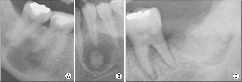
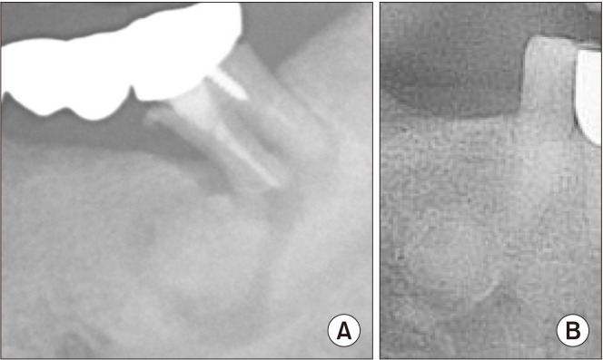
Results: COD was more prevalent in female patients. With the exception of three cases, all were focal COD. The majority of patients presented with symptoms when the lesion was smaller than 1.5 cm in size. Symptoms were observed when the apex of the tooth was included in the lesion or there was a local infection around the lesion. The history of tooth extraction and previous endodontic treatment were evaluated, and history was not a significant predictor for the onset of symptoms.
Conclusion: In this study, risk factors associated with symptomatic patients were size of lesion, apical involvement, and local infection.
Keywords: Cemento-osseous dysplasia; Florid cemento-osseous dysplasia; Focal cemento-osseoous dysplasia; Periapical cemental dysplasia.
Conflict of interest statement
No potential conflict of interest relevant to this article was reported.
Figures
Similar articles
-
Cemento-Osseous Dysplasia of the Jaw: Demographic and Clinical Analysis of 191 New Cases.Dent J (Basel). 2023 May 19;11(5):138. doi: 10.3390/dj11050138. Dent J (Basel). 2023. PMID: 37232789 Free PMC article.
-
Difficulties in the diagnosis of periapical translucencies and in the classification of cemento-osseous dysplasia.BMC Oral Health. 2019 Jul 10;19(1):139. doi: 10.1186/s12903-019-0843-0. BMC Oral Health. 2019. PMID: 31291935 Free PMC article.
-
Expansive focal cemento-osseous dysplasia.J Contemp Dent Pract. 2012 Jan 1;13(1):115-8. doi: 10.5005/jp-journals-10024-1105. J Contemp Dent Pract. 2012. PMID: 22430704
-
Familial florid cemento-osseous dysplasia: a report of three cases and review of the literature.Dentomaxillofac Radiol. 2021 Jan 1;50(1):20190486. doi: 10.1259/dmfr.20190486. Epub 2020 Apr 21. Dentomaxillofac Radiol. 2021. PMID: 32315206 Free PMC article. Review.
-
Periapical cemento-osseous dysplasia: a case report with twelve-year follow-up and review of literature.Int Endod J. 2015 Nov;48(11):1086-99. doi: 10.1111/iej.12417. Epub 2014 Dec 24. Int Endod J. 2015. PMID: 25425097 Review.
Cited by
-
Case report: Radiopaque mandibular lesions in three dogs.Front Vet Sci. 2025 Jan 15;11:1529669. doi: 10.3389/fvets.2024.1529669. eCollection 2024. Front Vet Sci. 2025. PMID: 39881726 Free PMC article.
-
Radiolucent lesions that may resemble inflammatory periapical lesions: A review article.Saudi Dent J. 2023 Dec;35(8):916-919. doi: 10.1016/j.sdentj.2023.11.003. Epub 2023 Nov 7. Saudi Dent J. 2023. PMID: 38107039 Free PMC article. Review.
-
The role of imaging in endodontics.Br Dent J. 2025 Apr;238(7):448-457. doi: 10.1038/s41415-025-8511-z. Epub 2025 Apr 11. Br Dent J. 2025. PMID: 40217027 Free PMC article. Review.
-
Craniofacial disorders and dysplasias: Molecular, clinical, and management perspectives.Bone Rep. 2024 Mar 1;20:101747. doi: 10.1016/j.bonr.2024.101747. eCollection 2024 Mar. Bone Rep. 2024. PMID: 38566929 Free PMC article. Review.
-
Periapical Cemento-Osseous Dysplasia in a Medically Compromised Patient: A Case Report.Cureus. 2023 May 29;15(5):e39623. doi: 10.7759/cureus.39623. eCollection 2023 May. Cureus. 2023. PMID: 37388605 Free PMC article.
References
-
- Alsufyani NA, Lam EW. Osseous (cemento-osseous) dysplasia of the jaws: clinical and radiographic analysis. J Can Dent Assoc. 2011;77:b70. - PubMed
-
- Mainville GN, Turgeon DP, Kauzman A. Diagnosis and management of benign fibro-osseous lesions of the jaws: a current review for the dental clinician. Oral Dis. 2017;23:440–50. doi: 10.1111/odi.12531. https://doi.org/10.1111/odi.12531. - DOI - PubMed
-
- MacDonald DS. Classification and nomenclature of fibro-osseous lesions. Oral Surg Oral Med Oral Pathol Oral Radiol. 2021;131:385–9. doi: 10.1016/j.oooo.2020.12.004. https://doi.org/10.1016/j.oooo.2020.12.004. - DOI - PubMed
-
- de Noronha Santos Netto J, Machado Cerri J, Miranda AM, Pires FR. Benign fibro-osseous lesions: clinicopathologic features from 143 cases diagnosed in an oral diagnosis setting. Oral Surg Oral Med Oral Pathol Oral Radiol. 2013;115:e56–65. doi: 10.1016/j.oooo.2012.05.022. https://doi.org/10.1016/j.oooo.2012.05.022. - DOI - PubMed
-
- Kim NK, Kim HS, Kim J, Nam W, Cha IH, Kim HJ. Florid cemento-osseous dysplasia: a report of two cases. J Korean Assoc Oral Maxillofac Surg. 2011;37:515–9. doi: 10.5125/jkaoms.2011.37.6.515. https://doi.org/10.5125/jkaoms.2011.37.6.515. - DOI - PubMed
LinkOut - more resources
Full Text Sources
Miscellaneous