Review
. 2022 Sep;32(9):773-785.
doi: 10.1016/j.tcb.2022.03.010. Epub 2022 Apr 29.

Mechanical regulation of cell-cycle progression and division

Affiliations
Review

Mechanical regulation of cell-cycle progression and division

Vivek K Gupta et al. Trends Cell Biol. 2022 Sep.

Abstract

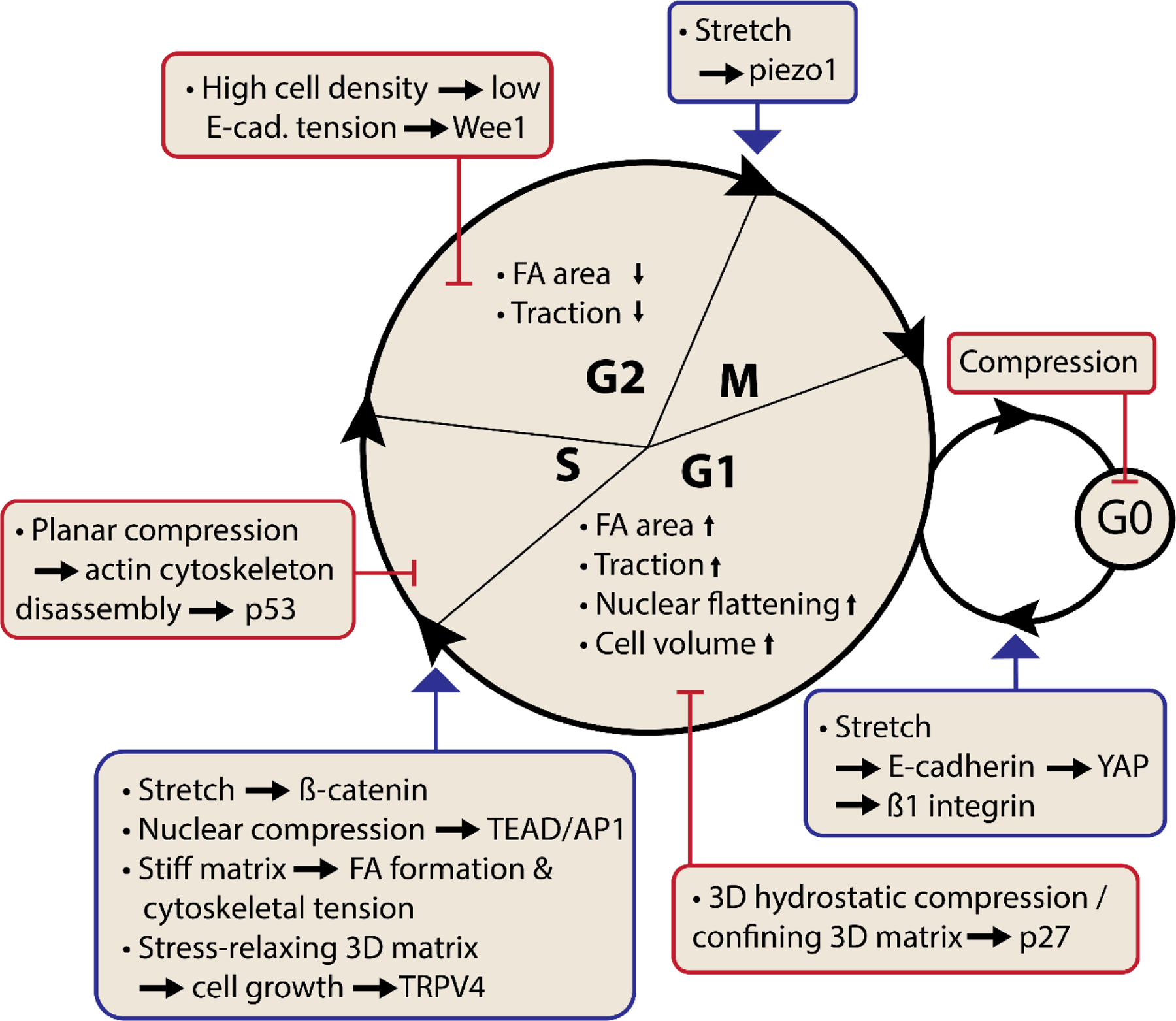
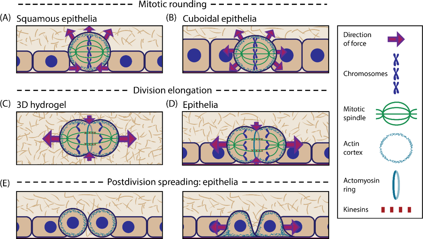
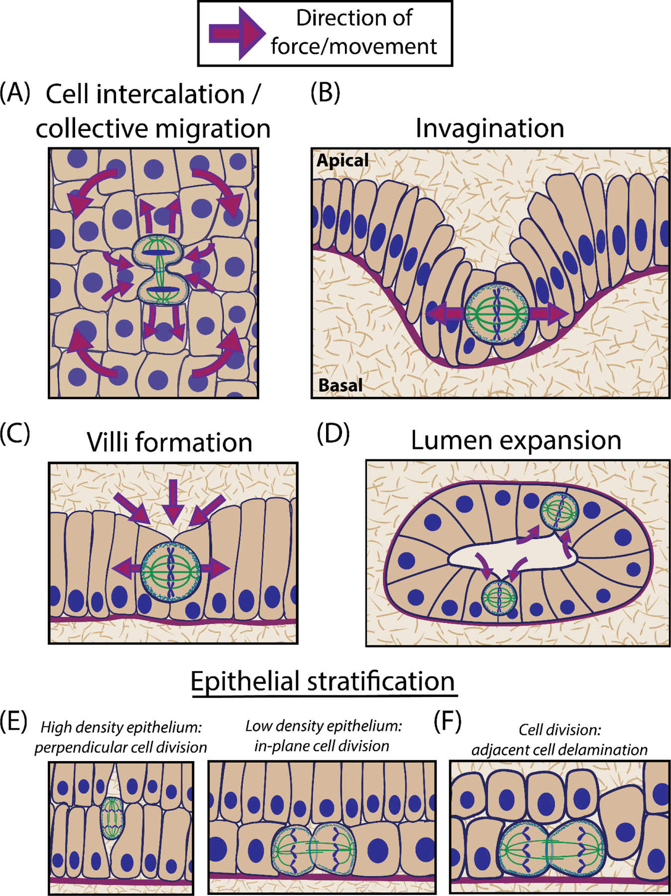
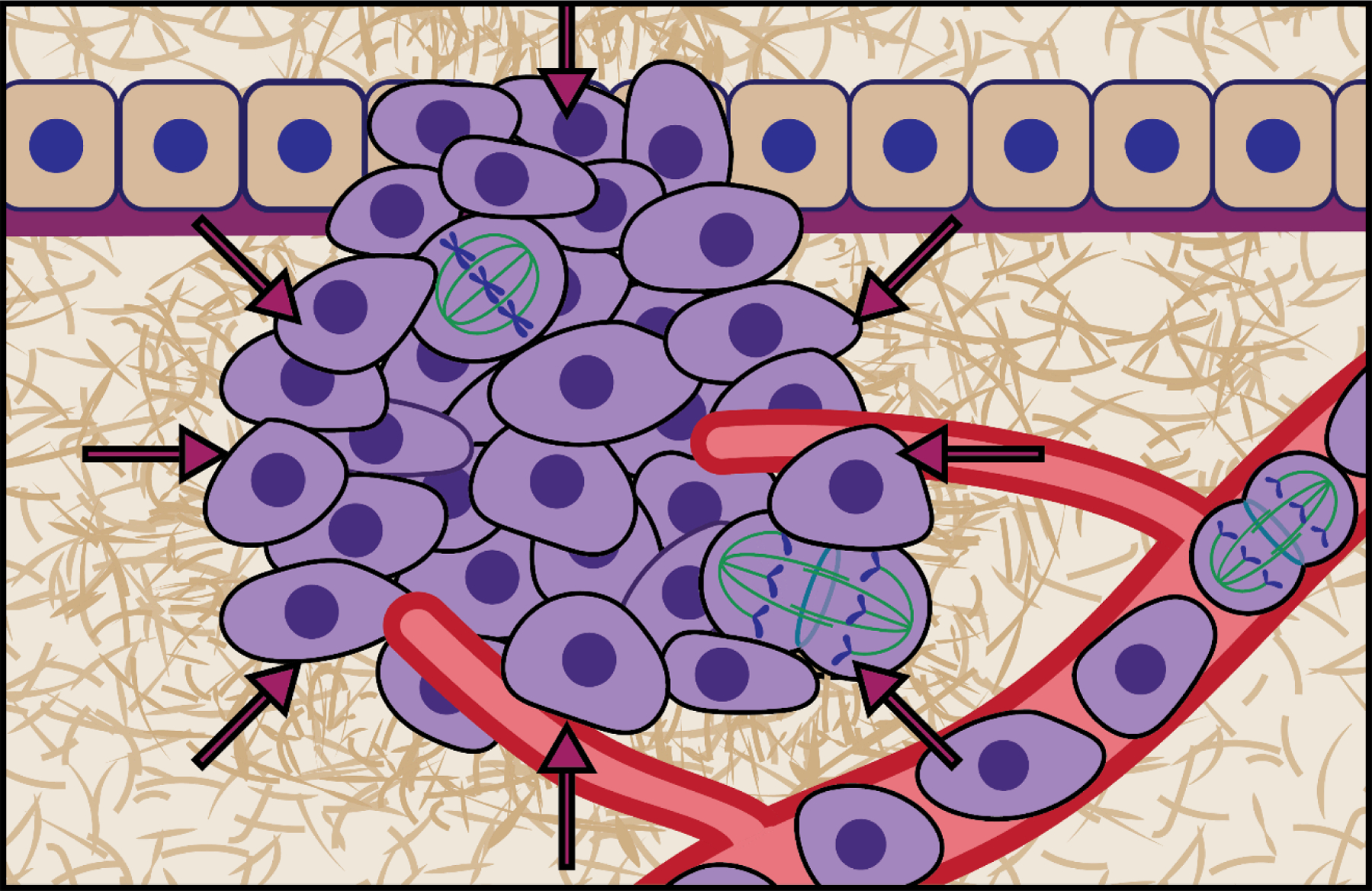
Cell-cycle progression and division are fundamental biological processes in animal cells, and their biochemical regulation has been extensively studied. An emerging body of work has revealed how mechanical interactions of cells with their microenvironment in tissues, including with the extracellular matrix (ECM) and neighboring cells, also plays a crucial role in regulating cell-cycle progression and division. We review recent work on how cells interpret physical cues and alter their mechanics to promote cell-cycle progression and initiate cell division, and then on how dividing cells generate forces on their surrounding microenvironment to successfully divide. Finally, the article ends by discussing how force generation during division potentially contributes to larger tissue-scale processes involved in development and homeostasis.

Keywords: cell cycle; cell division; extracellular matrix; force generation; mechanotransduction; microenvironment; mitosis.


Conflict of interest statement

Declaration of interests The authors declare no conflicts of interest.

Figures

Figure 1.
Figure 1.
Mechanical Regulation of Cell cycle Progression. Summary of recent work on how tension, compression, and extracellular matrix mechanics regulate cell cycle progression at various stages. Changes in cell properties are denoted within the circle, and applied mechanical perturbations, with their corresponding effect on cell cycle progression (blue arrows) or inhibition (red lines), are shown outside. Tissue tension can promote G0-G1 transition through β1 integrin signaling and YAP activation, G1-S transition through β-catenin, and G2-M transition through activation of piezo1. Similarly, intracellular tension (contractility) and spreading promotes G1-S transition. Conversely, compression inhibits cell cycle progression, in part due to the lack of activation of tension-dependent proliferation pathways. Changes in matrix mechanical properties including increased stiffness and increased stress relaxation, in 3D culture, can promote cell cycle progression through promotion of spreading and contractility and activation of a TRPV4-PI3K-p27 signaling axis, respectively. Note: relations depicted are dependent on biological context.
Figure 2.
Figure 2.
Extracellular Force Generation During Division. (A) During mitotic rounding, cells in flatter epithelia reduce their cross-sectional area within the monolayer plane, generating inward forces. (B) In more cuboidal epithelia, rounding leads to outward forces. (C, D) During elongation, cells generate outward forces through a combination of interpolar spindle elongation, and cytokinetic ring contraction due to conservation of cell volume; observed in 3D hydrogels (C) and within epithelia (D). (E) As newly divided daughter cells return to their native morphology, they may further exert forces. In epithelia, postdivision spreading leads to outward force generation.
Figure 3.
Figure 3.
Mechanical Forces Link Cell Division to Tissue-Scale Processes. (A) Cell division induces the intercalation of neighboring cells in between the newly formed daughter cells, as the daughter cells often migrate away from each other along the division axis. Larger-scale cell movements develop, pushing away from the division point along the division axis, and towards the division point along the perpendicular axes. (B) Outward forces generated during mitotic rounding can aid in epithelial buckling, leading to invagination [60]. (C) During mitotic rounding, pulling forces exerted along the apical-basal axis can lead to apical invagination, which precedes and aids in the formation of villi adjacent to these points [62]. (D) Pulling forces along the apical-basal axis during rounding can also create more room in developing lumens [63]. (E) In some contexts, dividing cells modify their orientation in response to shape cues determined by epithelial density, resulting in perpendicular divisions in a high density epithelium, and the formation/maintenance of stratified epithelia, or planar divisions in a low density epithelium, replenishing cells at the basal layer. (F) Division can also change the stress landscape in neighboring cells, inducing delamination [65]. Note: processes depicted are dependent on context and schematics are adapted from cited sources.
Figure I.
Figure I.
Cancer cells grow and divide in an increasingly confining environment due to increased solid stress, a stiffer ECM, and confinement due to the vasculature. Adapted from [39].

Similar articles

Cited by

References

    1. Finegan TM and Bergstralh DT (2019) Division orientation: disentangling shape and mechanical forces. Cell Cycle 18, 1187–1198 - PMC - PubMed
    1. Morgan DO (2007) Cell Cycle: Principles of Control, New Science Press.
    1. Gudipaty SA et al. (2017) Mechanical stretch triggers rapid epithelial cell division through Piezo1. Nature 543, 118–121 - PMC - PubMed
    1. Liang X et al. (2016) Mechanical stretching promotes skin tissue regeneration via enhancing mesenchymal stem cell homing and transdifferentiation. Stem Cells Transl. Med 5, 960–969 - PMC - PubMed
    1. Benham-Pyle BW et al. (2015) Mechanical strain induces E-cadherin-dependent Yap1 and β-catenin activation to drive cell cycle entry. Science 348, 1024–1027 - PMC - PubMed

Publication types