Review
. 2022 Nov;32(11):962-974.
doi: 10.1016/j.tcb.2022.03.011. Epub 2022 Apr 28.

A novel superfamily of bridge-like lipid transfer proteins

Affiliations
Review

A novel superfamily of bridge-like lipid transfer proteins

Sarah D Neuman et al. Trends Cell Biol. 2022 Nov.

Abstract

Lipid transfer proteins mediate nonvesicular transport of lipids at membrane contact sites to regulate the lipid composition of organelle membranes. Recently, a new type of bridge-like lipid transfer protein has emerged; these proteins contain a long hydrophobic groove and can mediate bulk transport of lipids between organelles. Here, we review recent insights into the structure of these proteins and identify a repeating modular unit that we propose to name the repeating β-groove (RBG) domain. This new structural understanding conceptually unifies all the RBG domain-containing lipid transfer proteins as members of an RBG protein superfamily. We also examine the biological functions of these lipid transporters in normal physiology and disease and speculate on the evolutionary origins of RBG proteins in bacteria.

Keywords: AlphaFold; Vps13; autophagy; lipid transfer proteins; lipids; membrane contact sites.


Conflict of interest statement

Declaration of interests The authors declare no competing interests.

Figures

Figure 1.
Figure 1.. Long hydrophobic groove lipid transfer proteins share structural similarities.
Ribbon models (top) and cross-sections of sphere models (bottom) showing representative members of five protein families that consist of long bridge-like structures comprised primarily of β-sheets (labeled blue in ribbon models), which form a groove lined with hydrophobic residues (glycine, alanine, valine, leucine, isoleucine, proline, phenylalanine, methionine and tryptophan; labeled orange in sphere models). The origins of the models are S. cerevisiae Vps13, H. sapiens ATG2A, H. sapiens Hobbit (KIAA0100), S. cerevisiae Tweek (Csf1) and H. sapiens SHIP164. The SHIP164 rendering omits two long unstructured loops (residues 873–1170 and 1360–1464). PDB files for Vps13 and Csf1 were obtained from Jerry Yang and William Prinz, who used trRosetta to predict regions of ~1000 amino acids, then assembled a complete model from overlapping segments [16]. PDB files for ATG2A, Hob and SHIP164 were obtained from the AlphaFold database [21]. All renderings were generated in ChimeraX [78]. Note that the VPS13 model contains a disconnect in the hydrophobic groove coincident with a large insert called the VAB domain ([23]; further discussed in Fig. 3).
Figure 2.
Figure 2.. The “repeating β-groove” (RBG) domain is the modular building block of RBG lipid transfer proteins.
(A) Ribbon model of D. melanogaster Hobbit with RBG domains alternately labeled in pink, yellow and blue. Rendering generated using PyMOL version 2.3.4. PDB file obtained from the AlphaFold database [21]. (B) Model of a single RBG domain from D. melanogaster Hobbit showing the basic RBG domain structure consisting of five antiparallel β-strands (blue) that form a groove, followed by an unstructured loop that crosses back over the β-sheet (gray). Ribbon models: left – front view, middle – side-view (90° rotation). Note the presence of strand-breaking residues (colored pink) near the middle of each strand that generate curvature. (C) Sphere model side-view of the same RBG domain in (B), demonstrating that the concave surface (interior) of the RBG domain’s groove is lined with hydrophobic residues (orange), while the convex surface (exterior) is hydrophilic (gray). Strandbreaking residues shown in pink. Models generated with ChimeraX [78]. (D) Cartoon illustration of three contiguous RBG domains. The direction of each β-strand and loop is indicated by arrowheads. (E) Illustration of the number of RBG domains present in each RBG protein family, as well as the total length of each predicted protein. Note that the initial and final RBG domains are shorter in each protein (indicated by “−”); additionally, Tweek contains two RBG domains with three β-strands and one with seven β-strands (indicated by “+”).
Figure 3.
Figure 3.. Structural features of RBG proteins in eukaryotes and prokaryotes.
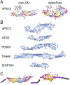
(A) Ribbon models of cryo-EM structure of Chaetomium thermophilum Vps13 (left; residues 6–1381) [9] and AlphaFold prediction of rat VPS13A (right; residues 1–1388) [21] show consistency of RBG domain structure between experimental and predicted models. Differences include diameter of the hydrophobic groove and degree of superhelical rotation. Coloring: RBG domains as Figure 2A; helical “handle” [9] gray. PDB file of Vps13 cryo-EM provided by Karin Reinisch [9]. (B) Ribbon models of the five eukaryotic RBG proteins showing only β-sheets; long loops and all helices omitted. (C) Ribbon models of YicH (E. coli) and Mdm31 (mitochondrial protein) highlight structural similarities with eukaryotic RBG proteins. Coloring: RBG domains as Figure 2A; terminal helices: purple; predicted transmembrane helices: black. Note adaptation of Mdm31 to integrate into both inner and outer mitochondrial membranes. PDB files for ATG2A (human), Hobbit (fly), SHIP164 (human), YicH (E. coli) and Mdm31 (yeast, omitting long unstructured loops) obtained from AlphaFold database [21]. Tweek model is of yeast Csf1, provided by Rosario Valentini [17]. VPS13 model in (B) was generated by submitting residues 1643–2111+2543–2840 from yeast Vps13 (omitting 67% of the VAB domain insert) to AlphaFold [60]. Resulting structure was overlapped with AlphaFold model of rat VPS13A (residues 1–2335). Other published Vps13 models ([16]; Fig. 1) show a disconnect in the hydrophobic groove where VAB domain (>700 residues; [23]) is inserted. Continuity traversing this insert, as shown here, is strongly predicted (probability local distance difference test (pLDDT) >0.9 [21]).

References

    1. Kaplan MR and Simoni RD (1985) Intracellular transport of phosphatidylcholine to the plasma membrane. J. Cell Biol 101, 441–445 - PMC - PubMed
    1. DeGrella RF and Simoni RD (1982) Intracellular transport of cholesterol to the plasma membrane. J. Biol. Chem 257, 14256–14262 - PubMed
    1. Wirtz KW and Zilversmit DB (1968) Exchange of phospholipids between liver mitochondria and microsomes in vitro. J. Biol. Chem 243, 3596–3602 - PubMed
    1. Wong LH et al. (2019) Lipid transfer proteins: the lipid commute via shuttles, bridges and tubes. Nat. Rev. Mol. Cell Biol 20, 85–101 - PubMed
    1. Wong LH et al. (2017) Advances on the Transfer of Lipids by Lipid Transfer Proteins. Trends Biochem. Sci 42, 516–530 - PMC - PubMed

Publication types