

Oleanolic acid administration alleviates neuropathic pain after a peripheral nerve injury by regulating microglia polarization-mediated neuroinflammation
- PMID: 35492085
- PMCID: PMC9051258
- DOI: 10.1039/c9ra10388k
Oleanolic acid administration alleviates neuropathic pain after a peripheral nerve injury by regulating microglia polarization-mediated neuroinflammation
Abstract
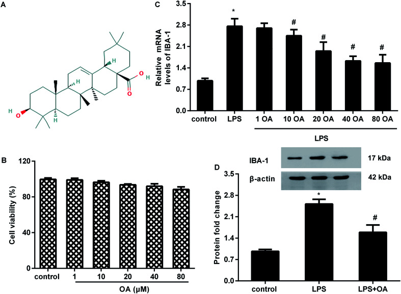
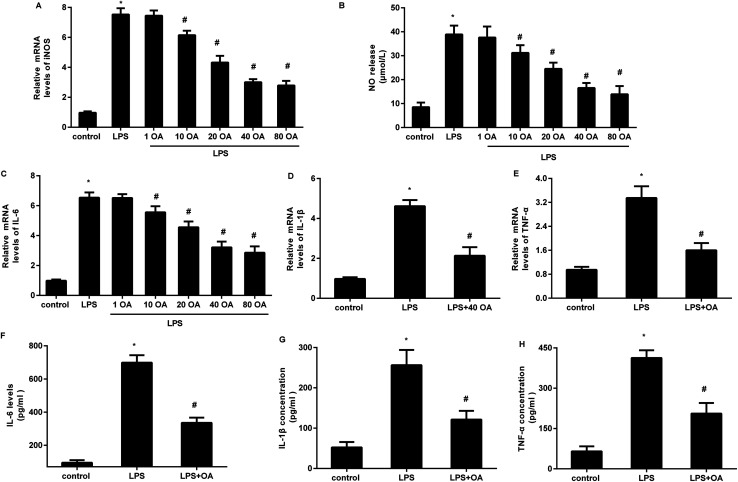
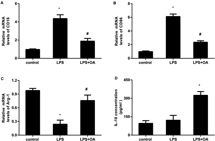
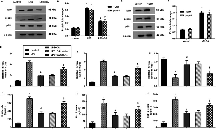
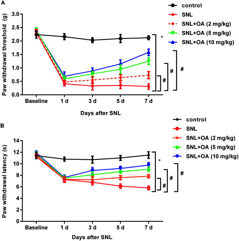
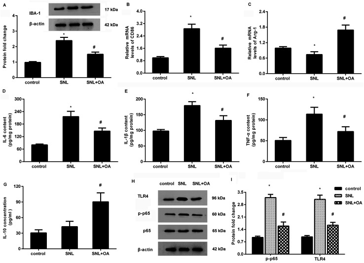
Neuropathic pain caused by a peripheral nerve injury constitutes a great challenge in clinical treatments due to the unsatisfactory efficacy of the current strategy. Microglial activation-mediated neuroinflammation is a major characteristic of neuropathic pain. Oleanolic acid is a natural triterpenoid in food and medical plants, and fulfills pleiotropic functions in inflammatory diseases. Nevertheless, its role in neuropathic pain remains poorly elucidated. In the current study, oleanolic acid dose-dependently suppressed LPS-evoked IBA-1 expression (a microglial marker) without cytotoxicity to microglia, suggesting the inhibitory efficacy of oleanolic acid in microglial activation. Moreover, oleanolic acid incubation offset LPS-induced increases in the iNOS transcript and NO releases from microglia, concomitant with the decreases in pro-inflammatory cytokine transcripts and production including IL-6, IL-1β, and TNF-α. Simultaneously, oleanolic acid shifted the microglial polarization from the M1 phenotype to the M2 phenotype upon LPS conditions by suppressing LPS-induced M1 marker CD16, CD86 transcripts, and enhancing the M2 marker Arg-1 mRNA and anti-inflammatory IL-10 levels. In addition, the LPS-induced activation of TLR4-NF-κB signaling was suppressed in the microglia after the oleanolic acid treatment. Restoring this signaling by the TLR4 plasmid transfection overturned the suppressive effects of oleanolic acid on microglial polarization-evoked inflammation. In vivo, oleanolic acid injection alleviated allodynia and hyperalgesia in SNL-induced neuropathic pain mice. Concomitantly, oleanolic acid facilitated microglial polarization to M2, accompanied by inhibition in inflammatory cytokine levels and activation of TLR4-NF-κB signaling. Collectively, these findings confirm that oleanolic acid may ameliorate neuropathic pain by promoting microglial polarization from pro-inflammatory M1 to anti-inflammatory M2 phenotype via the TLR4-NF-κB pathway, thereby indicating its usefulness as therapeutic intervention in neuropathic pain.
This journal is © The Royal Society of Chemistry.
Conflict of interest statement
There are no conflicts to declare.
Figures
Similar articles
-
Curcumin inhibits LPS-induced neuroinflammation by promoting microglial M2 polarization via TREM2/ TLR4/ NF-κB pathways in BV2 cells.Mol Immunol. 2019 Dec;116:29-37. doi: 10.1016/j.molimm.2019.09.020. Epub 2019 Oct 4. Mol Immunol. 2019. PMID: 31590042
-
( +)-Catechin Alleviates CCI-Induced Neuropathic Pain by Modulating Microglia M1 and M2 Polarization via the TLR4/MyD88/NF-κB Signaling Pathway.J Neuroimmune Pharmacol. 2025 Apr 7;20(1):33. doi: 10.1007/s11481-025-10202-9. J Neuroimmune Pharmacol. 2025. PMID: 40195186
-
Glycyrrhizin ameliorates inflammatory pain by inhibiting microglial activation-mediated inflammatory response via blockage of the HMGB1-TLR4-NF-kB pathway.Exp Cell Res. 2018 Aug 1;369(1):112-119. doi: 10.1016/j.yexcr.2018.05.012. Epub 2018 May 12. Exp Cell Res. 2018. PMID: 29763588
-
The natural (poly)phenols as modulators of microglia polarization via TLR4/NF-κB pathway exert anti-inflammatory activity in ischemic stroke.Eur J Pharmacol. 2022 Jan 5;914:174660. doi: 10.1016/j.ejphar.2021.174660. Epub 2021 Dec 1. Eur J Pharmacol. 2022. PMID: 34863710 Review.
-
Focus on P2X7R in microglia: its mechanism of action and therapeutic prospects in various neuropathic pain models.Front Pharmacol. 2025 Mar 25;16:1555732. doi: 10.3389/fphar.2025.1555732. eCollection 2025. Front Pharmacol. 2025. PMID: 40201695 Free PMC article. Review.
Cited by
-
β-Sitosterol Alleviates Neuropathic Pain by Affect Microglia Polarization through Inhibiting TLR4/NF-κB Signaling Pathway.J Neuroimmune Pharmacol. 2023 Dec;18(4):690-703. doi: 10.1007/s11481-023-10091-w. Epub 2023 Dec 2. J Neuroimmune Pharmacol. 2023. PMID: 38041701
-
Temporal changes of spinal microglia in murine models of neuropathic pain: a scoping review.Front Immunol. 2024 Dec 6;15:1460072. doi: 10.3389/fimmu.2024.1460072. eCollection 2024. Front Immunol. 2024. PMID: 39735541 Free PMC article.
-
From Plant to Chemistry: Sources of Active Opioid Antinociceptive Principles for Medicinal Chemistry and Drug Design.Molecules. 2023 Oct 14;28(20):7089. doi: 10.3390/molecules28207089. Molecules. 2023. PMID: 37894567 Free PMC article. Review.
-
Effects of Natural Product-Derived Compounds on Inflammatory Pain via Regulation of Microglial Activation.Pharmaceuticals (Basel). 2023 Jun 29;16(7):941. doi: 10.3390/ph16070941. Pharmaceuticals (Basel). 2023. PMID: 37513853 Free PMC article. Review.
-
Netrin-1 Promotes M2 Type Activation and Inhibits Pyroptosis of Microglial Cells by Depressing RAC1/Nf-?B Pathway to Alleviate Inflammatory Pain.Physiol Res. 2024 Apr 30;73(2):305-314. doi: 10.33549/physiolres.935134. Physiol Res. 2024. PMID: 38710054 Free PMC article.
References
LinkOut - more resources
Full Text Sources
Research Materials
Miscellaneous