. 2022 May 20;25(5):104293.
doi: 10.1016/j.isci.2022.104293. Epub 2022 Apr 25.

Inhibitors of dihydroorotate dehydrogenase cooperate with molnupiravir and N4-hydroxycytidine to suppress SARS-CoV-2 replication

Affiliations

Inhibitors of dihydroorotate dehydrogenase cooperate with molnupiravir and N4-hydroxycytidine to suppress SARS-CoV-2 replication

Kim M Stegmann et al. iScience. .

Abstract

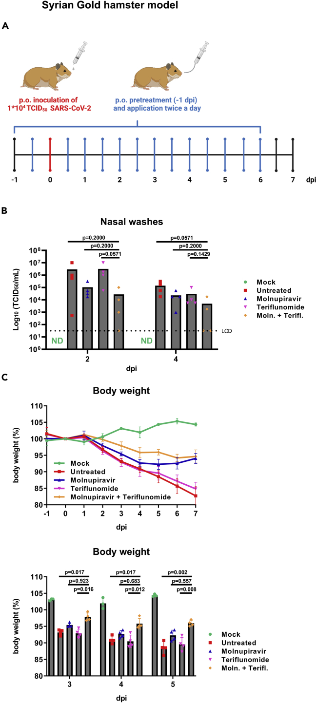
The nucleoside analog N4-hydroxycytidine (NHC) is the active metabolite of the prodrug molnupiravir, which has been approved for the treatment of COVID-19. SARS-CoV-2 incorporates NHC into its RNA, resulting in defective virus genomes. Likewise, inhibitors of dihydroorotate dehydrogenase (DHODH) reduce virus yield upon infection, by suppressing the cellular synthesis of pyrimidines. Here, we show that NHC and DHODH inhibitors strongly synergize in the inhibition of SARS-CoV-2 replication in vitro. We propose that the lack of available pyrimidine nucleotides upon DHODH inhibition increases the incorporation of NHC into nascent viral RNA. This concept is supported by the rescue of virus replication upon addition of pyrimidine nucleosides to the media. DHODH inhibitors increased the antiviral efficiency of molnupiravir not only in organoids of human lung, but also in Syrian Gold hamsters and in K18-hACE2 mice. Combining molnupiravir with DHODH inhibitors may thus improve available therapy options for COVID-19.

Keywords: Drugs; Virology.


Conflict of interest statement

AS, HK, EP, and DV are employees of Immunic AG and own shares and/or stock-options of the parent company of Immunic AG, Immunic Inc. Some of the Immunic AG employees also hold patents for the Immunic compounds described in this manuscript (WO2012/001,148, WO03006425). KMS, AD, and MD are employees of University Medical Center Göttingen, which has signed a License Agreement with Immunic AG covering the combination of DHODH inhibitors and nucleoside analogs to treat viral infections, including COVID-19 (inventors: MD, KMS, and AD). The other authors declare no conflict of interest.

Figures

None
Graphical abstract
Figure 1
Figure 1
The combination of N4-hydroxycytidine (NHC) and inhibitors of dihydroorotate dehydrogenase (DHODH) strongly impairs SARS-CoV-2 replication without detectable cytotoxicity (A) Mechanistic concept for the synergistic inhibitory effect of DHODH inhibitors and N4-hydroxycytidine (NHC) on SARS-CoV-2 RNA replication. Biosynthesis of pyrimidines starts with carbamoyl phosphate and aspartate to form dihydroorotate. Dihydroorotate is further oxidized to orotate by dihydroorotate dehydrogenase (DHODH) and later converted to uridine triphosphate (UTP) and cytidine triphosphate (CTP). Molnupiravir is the prodrug of NHC, which is further converted to the corresponding triphosphate (NHCTP), which competes with CTP for incorporation into nascent virus RNA. The suppression of CTP synthesis by inhibitors of DHODH is expected to enhance the incorporation of NHCTP into the viral RNA, causing false incorporation of nucleotides in subsequent rounds of replication. (B) Reduced cytopathic effect (CPE) by NHC and DHODH inhibitors. Vero E6 cells were treated with drugs or the DMSO control for 24 h, inoculated with SARS-CoV-2 (strain GOE_001), and further incubated in the presence of the same drugs for 48 h. Cell morphology was assessed by phase contrast microscopy. Note that the CPE was readily visible in virus-infected cells, in DMSO-treated cells and also when cells had been treated with either drug alone. However, the CPE was observed only to a far lesser extent when the cells had been incubated with both NHC and DHODH inhibitors. Bar, 100 μm. (C) Reduction of the median tissue culture infectious dose (TCID50) by the combination of NHC and the DHODH inhibitor IMU-838. Vero E6 cells were treated with NHC, IMU-838, or the combination of both compounds for 24 h before infection, and then throughout the time of infection. Cells were infected with SARS-CoV-2, strain hCoV-19/Germany/BY-Bochum-1/2020 (MOI 0.1), and further incubated for 48 h. The supernatant was titrated to determine the TCID50/mL (mean with SD, n = 3; logarithmic scale). (D) Lack of measurable cytotoxicity by NHC and DHODH inhibitors. Vero E6 cells were treated with NHC and/or IMU-838, BAY2402234, and teriflunomide at the indicated concentrations for 72 h. The release of lactate dehydrogenase (LDH) to the supernatant was quantified by bioluminescence as a readout for cytotoxicity. The percentages reflect the proportion of LDH released to the media, compared to the overall amount of LDH in the cells (LDH control) (mean with SD, n = 3).
Figure 2
Figure 2
Strong synergism of NHC and DHODH inhibitors to diminish the release of SARS-CoV-2 RNA from cultured cells (A) Reduced release of viral RNA upon combined treatment with NHC and IMU-838. Vero E6 cells were treated with NHC and/or IMU-838, and infected as in Figure 1. A sample of the inoculum was preserved for RNA preparation. At 48 h post infection (p.i.), RNA was isolated from the cell supernatants, followed by quantitative RT-PCR to detect viral RNA and determine the amount of SARS-CoV-2 RNA copies per mL (mean, n = 3). The synergy score was calculated using the Bliss independence model. Data are presented as mean ± SEM. A Bliss score >10 is generally considered to reveal strong drug synergism. (B) Diminished virus RNA progeny by NHC and DHODH inhibitors even when added 4 h after SARS-CoV-2 infection. Vero E6 cells were infected as described in Figure 1 and treated with NHC and/or DHODH inhibitors at 4 h post infection (p.i.). RNA was isolated from the cell supernatants, and SARS-CoV-2 RNA was quantified by qRT-PCR. The amount of RNA found upon infection without drug treatment was defined as 100%, and the other RNA quantities were normalized accordingly. RNA was also isolated from the virus inoculum used to infect the cells. Note that the combination treatment reduced SARS-CoV-2 replication to a greater extent compared to single drug treatments even when applied 4 h p.i. (mean with SD, n = 3). For p values, see Figure S2C. (C) Reduced virus RNA progeny in the presence of NHC and various DHODH inhibitors. Vero E6 cells were treated with drugs and/or infected as in Figure 1, followed by quantitative detection of SARS-CoV-2 RNA. The drug combinations were found capable of reducing virus RNA yield by more than 100-fold as compared to single drug treatments (mean with SD, n = 3). (D) In Calu-3 cells, too, the combination of NHC and the DHODH inhibitors IMU-838, BAY2402234, or teriflunomide strongly reduced the amount of viral RNA released to the supernatant (mean with SD, n = 3).
Figure 3
Figure 3
Synergistic reduction of viral protein synthesis by NHC and DHODH inhibitors (A–C) Representative images showing the reduction of viral protein synthesis by NHC and BAY2402234 (A), teriflunomide (B), or IMU-838 (C). Vero E6 cells were treated and infected with SARS-CoV-2 as in Figure 1. Cell nuclei were stained with 4′,6-Diamidino-2-phenylindole (DAPI), and the SARS-CoV-2 spike and nucleoprotein were detected by immunofluorescence. Bar, 100 μm. (D–F) Reduced viral protein synthesis in the presence of NHC and DHODH inhibitors. Upon drug treatment and/or infection of Vero E6 cells as in Figure 1, the viral spike and nucleoprotein as well as DHODH and HSC70 (loading control) were detected by immunoblot analysis. The absence of a signal corresponding to spike or nucleoprotein in non-infected samples ensures the specificity of the antibodies for virus proteins.
Figure 4
Figure 4
The combination of NHC with DHODH inhibitors synergistically reduces the replication of SARS-CoV-2 variants in Vero E6 cells (A–D) The combination of NHC with IMU-838 synergistically reduced the progeny of SARS-CoV-2 variants. The TCID50 of virus progeny was determined upon treatment with NHC and DHODH inhibitors, and infection with the original SARS-CoV-2 or the SARS-CoV-2 variants Alpha, Beta, and Delta. Vero E6 cells were treated with drugs for 24 h before and then throughout the infection. Cells were infected with original SARS-CoV-2 (hCoV-19/Germany/BY-Bochum-1/2020, (A)), Alpha (B.1.1.7, (B)) Beta (B.1.351, (C)), or Delta (B.1.617.2, (D)) (MOI 0.1) and further incubated for 48 h. The supernatant was titrated to determine the TCID50/mL. Note that all variants responded similarly to the original strain, indicating that the drug combination is effective against SARS-CoV-2 variants.
Figure 5
Figure 5
Uridine as well as cytidine rescue SARS-CoV-2 replication in the presence of NHC and DHODH inhibitors (A) The antiviral effect of DHODH inhibitors combined with NHC can be reverted by uridine. Vero E6 cells were treated with drugs and inoculated with SARS-CoV-2 as in Figure 1. On top of the drugs, where indicated, uridine was added to the cell culture media, at concentrations of 2, 5, or 10 μM. SARS-CoV-2 propagation was still diminished by NHC and DHODH inhibitors despite 2 μM uridine levels, but rescued in the presence of 5 or 10 μM uridine (mean with SD, n = 3), in agreement with the mechanism outlined in Figure 1A. For p values, see Figure S4A. (B) Restored SARS-CoV-2 replication by cytidine, in the presence of NHC and DHODH inhibitors. The experiment was carried out as in (A), with the addition of cytidine instead of uridine. 5 or 10 μM cytidine restored virus replication in the presence of the drugs (mean with SD, n = 3), further confirming the mechanism outlined in Figure 1A. For p values, see Figure S4B.
Figure 6
Figure 6
Reduced SARS-CoV-2 propagation and dsRNA formation by NHC and BAY2402234 in human lung organoids (A) Reduced TCID50 by NHC and the DHODH inhibitor BAY2402234. Human stem cell-derived lung organoids were sliced and treated with 1 μM NHC and/or 1 μM BAY2402234 for 24 h before and then throughout the time of infection. Organoid slices were infected with 35,000 PFU per well and further incubated for 24, 48, or 72 h. The supernatant was titrated to determine the TCID50/mL (mean with SD, n = 6). Statistical evaluation was performed using the Mann-Whitney U test. (B) Cell viability of lung organoids was not detectably affected by NHC and/or the DHODH inhibitor BAY2402234. The release of lactate dehydrogenase (LDH) to the supernatant was quantified by bioluminescence as a readout for cytotoxicity and cell viability as in Figure 1D (mean with SD, n = 6). (C) Representative images showing the reduction of viral double-stranded RNA (dsRNA) formation in lung organoid cells upon treatment with NHC and BAY2402234. Human lung organoid slices were treated and infected as in (A). For immunofluorescence analysis, samples were permeabilized and subjected to staining with an antibody against dsRNA (green dots within cytoplasms). Cell nuclei were stained with Hoechst33342. Bar, 10 μm.
Figure 7
Figure 7
DHODH inhibitors cooperate with Molnupiravir for treating COVID-19 in Syrian Gold hamsters (A) Treatment and infection scheme (drawn with BioRender.com). Male Syrian Gold hamsters (n = 4) were treated with 250 mg/kg molnupiravir alone, 10 mg/kg teriflunomide alone, or a combination of both, administered twice a day, starting 24 h before inoculation until six days post inoculation with 1∗104 TCID50 SARS-CoV-2. (B) Virus load within nasal washes, determined by TCID50. Nasal washes were obtained at days 2 and 4 post infection (p.i.), and titrated to determine the content of virus. On average, virus load was reduced by 1–2 orders of magnitude as compared to non-treated animals, and the drug combination yielded the strongest reduction. Statistical evaluation was performed using the Mann-Whitney U test (mean with SD, n = 4). (C) Decreased loss in body weight of SARS-CoV-2-infected hamsters in the presence of molnupiravir and teriflunomide. Hamsters were treated and infected as in A, and body weights were documented daily for seven days. Data points represent the body weight of each animal (% of weight at day 0, mean with SD, n = 4). Statistical analysis was performed by one-way ANOVA followed by post hoc Tukey tests (p < 0.05) to reveal that the drug combination was statistically more effective than single drugs on days 3 through 5 post infection. For p values, see Figure S5C.
Figure 8
Figure 8
DHODH inhibitors cooperate with molnupiravir for treating COVID-19 in K18-hACE-2 mice (A) Treatment and infection scheme (drawn with BioRender.com). Female K18-hACE-2 mice (n = 8) received either 50 mg/kg bid molnupiravir, 10 mg/kg/day teriflunomide, or a mixture of molnupiravir + teriflunomide via oral gavage 2 h prior to infection with SARS-CoV-2, and 6 h thereafter. The drugs were then provided to the animals in a 12 h application cycle. The untreated control group received the vehicle solution PEG400. (B) Reduced SARS-CoV-2 RNA load in the lungs of mice upon combinatory treatment with molnupiravir and DHODH inhibitors. Viral RNA was isolated from lung homogenates and quantified by qRT-PCR 4 days after infection. Data points represent the viral RNA copy number found for each animal, along with the geometric mean of each group (n = 8). Reduction in viral load is shown as fold reduction compared to the untreated control. Statistical evaluation was performed using the Mann-Whitney U test. For p values, see Figure S7B. (C) Reduced lymphocyte infiltration of the lungs of SARS-CoV-2-infected mice upon treatment with molnupiravir and DHODH inhibitors. Female K18-hACE-2 mice (n = 8) were treated and infected as in (A), followed by histopathological analysis of lungs (H&E stains). For comparison, lungs from non-infected mice of the same genotype, taken from a different series of experiments, were investigated. The infiltration of the lungs with perivascular and interstitial lymphocytes was first scored separately (0–3), and scores were summed up for each animal. The scores (points) for each animal are depicted, along with the median of each group (n = 8). Statistical evaluation was performed by the Mann-Whitney U test. For p values, see Figure S7C. (D) Representative images of lymphocyte infiltration, as described in (C). Images of non-infected lung tissue from K18-hACE-2 mice were taken from a different series (historical control) and added to the panel. Bar, 100 μm.

References

    1. Abdelnabi R., Foo C.S., De Jonghe S., Maes P., Weynand B., Neyts J. Molnupiravir inhibits replication of the emerging SARS-CoV-2 variants of concern in a hamster infection model. J. Infect. Dis. 2021;224:749–753. doi: 10.1093/infdis/jiab361. - DOI - PMC - PubMed
    1. Agostini M.L., Pruijssers A.J., Chappell J.D., Gribble J., Lu X., Andres E.L., Bluemling G.R., Lockwood M.A., Sheahan T.P., Sims A.C., et al. Small-molecule antiviral β-d-N (4)-hydroxycytidine inhibits a proofreading-intact coronavirus with a high genetic barrier to resistance. J. Virol. 2019;93:e01348-19. doi: 10.1128/jvi.01348-19. - DOI - PMC - PubMed
    1. Barnard D.L., Hubbard V.D., Burton J., Smee D.F., Morrey J.D., Otto M.J., Sidwell R.W. Inhibition of severe acute respiratory syndrome-associated coronavirus (SARSCoV) by calpain inhibitors and beta-D-N4-hydroxycytidine. Antivir. Chem. Chemother. 2004;15:15–22. doi: 10.1177/095632020401500102. - DOI - PubMed
    1. Beigel J.H., Tomashek K.M., Dodd L.E., Mehta A.K., Zingman B.S., Kalil A.C., Hohmann E., Chu H.Y., Luetkemeyer A., Kline S., et al. Remdesivir for the treatment of COVID-19 - final report. N. Engl. J. Med. 2020;383:1813–1826. doi: 10.1056/nejmoa2007764. - DOI - PMC - PubMed
    1. Bliss C.I. The toxicity of poisons applied jointly. Ann. Appl. Biol. 1939;26:585–615. doi: 10.1111/j.1744-7348.1939.tb06990.x. - DOI