

The Role of the Melatoninergic System in Circadian and Seasonal Rhythms-Insights From Different Mouse Strains
- PMID: 35492605
- PMCID: PMC9039042
- DOI: 10.3389/fphys.2022.883637
The Role of the Melatoninergic System in Circadian and Seasonal Rhythms-Insights From Different Mouse Strains
Abstract
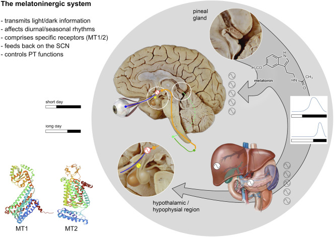
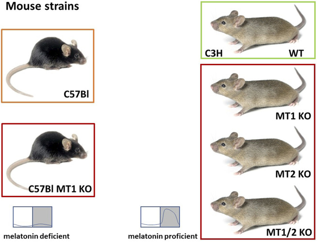
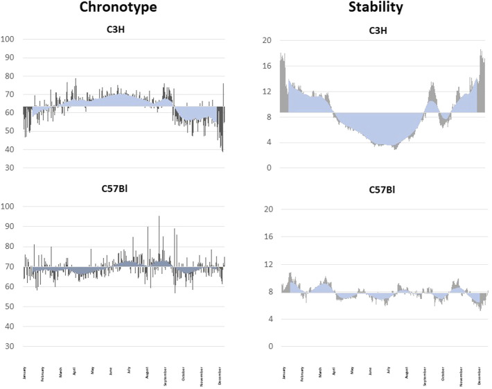
The melatoninergic system comprises the neurohormone melatonin and its molecular targets. The major source of melatonin is the pineal organ where melatonin is rhythmically produced during darkness. In mammals, melatonin biosynthesis is controlled by the central circadian rhythm generator in the suprachiasmatic nucleus (SCN) and photoreceptors in the retina. Melatonin elicits its function principally through two specific receptors called MT1 and MT2. MT1 is highly expressed in the SCN and the hypophysial pars tuberalis (PT), an important interface for control of seasonal functions. The expression of the MT2 is more widespread. The role of the melatoninergic system in the control of seasonal functions, such as reproduction, has been known for more than 4 decades, but investigations on its impact on the circadian system under normal (entrained) conditions started 2 decades later by comparing mouse strains with a fully functional melatoninergic system with mouse strains which either produce insufficient amounts of melatonin or lack the melatonin receptors MT1 and MT2. These studies revealed that an intact melatoninergic system is not required for the generation or maintenance of rhythmic behavior under physiological entrained conditions. As shown by jet lag experiments, the melatoninergic system facilitated faster re-entrainment of locomotor activity accompanied by a more rapid adaptation of the molecular clock work in the SCN. This action depended on MT2. Further studies indicated that the endogenous melatoninergic system stabilizes the locomotor activity under entrained conditions. Notably, these effects of the endogenous melatoninergic system are subtle, suggesting that other signals such as corticosterone or temperature contribute to the synchronization of locomotor activity. Outdoor experiments lasting for a whole year indicate a seasonal plasticity of the chronotype which depends on the melatoninergic system. The comparison between mice with an intact or a compromised melatoninergic system also points toward an impact of this system on sleep, memory and metabolism.
Keywords: activity rhythms; chronobiology; jet lag; melatonin; mouse strains; seasonality.
Copyright © 2022 Pfeffer, von Gall, Wicht and Korf.
Conflict of interest statement
The authors declare that the research was conducted in the absence of any commercial or financial relationships that could be construed as a potential conflict of interest.
Figures
Similar articles
-
Mouse Models in Circadian Rhythm and Melatonin Research.J Pineal Res. 2024 Aug;76(5):e12986. doi: 10.1111/jpi.12986. J Pineal Res. 2024. PMID: 38965880 Review.
-
The Role of the Melatoninergic System in Light-Entrained Behavior of Mice.Int J Mol Sci. 2017 Mar 1;18(3):530. doi: 10.3390/ijms18030530. Int J Mol Sci. 2017. PMID: 28257037 Free PMC article.
-
The endogenous melatonin (MT) signal facilitates reentrainment of the circadian system to light-induced phase advances by acting upon MT2 receptors.Chronobiol Int. 2012 May;29(4):415-29. doi: 10.3109/07420528.2012.667859. Epub 2012 Apr 10. Chronobiol Int. 2012. PMID: 22489607
-
Synchronizing effects of melatonin on diurnal and circadian rhythms.Gen Comp Endocrinol. 2018 Mar 1;258:215-221. doi: 10.1016/j.ygcen.2017.05.013. Epub 2017 May 19. Gen Comp Endocrinol. 2018. PMID: 28533170 Review.
-
Signaling pathways to and from the hypophysial pars tuberalis, an important center for the control of seasonal rhythms.Gen Comp Endocrinol. 2018 Mar 1;258:236-243. doi: 10.1016/j.ygcen.2017.05.011. Epub 2017 May 13. Gen Comp Endocrinol. 2018. PMID: 28511899 Review.
Cited by
-
Melatonin/Sericin Wound Healing Patches: Implications for Melanoma Therapy.Int J Mol Sci. 2024 Apr 29;25(9):4858. doi: 10.3390/ijms25094858. Int J Mol Sci. 2024. PMID: 38732075 Free PMC article. Review.
-
Mediterranean Diet and Melatonin: A Systematic Review.Antioxidants (Basel). 2023 Jan 24;12(2):264. doi: 10.3390/antiox12020264. Antioxidants (Basel). 2023. PMID: 36829823 Free PMC article. Review.
-
Melanogenesis Is Directly Affected by Metabolites of Melatonin in Human Melanoma Cells.Int J Mol Sci. 2023 Oct 6;24(19):14947. doi: 10.3390/ijms241914947. Int J Mol Sci. 2023. PMID: 37834395 Free PMC article.
-
One seasonal clock fits all?J Comp Physiol A Neuroethol Sens Neural Behav Physiol. 2024 Jul;210(4):641-647. doi: 10.1007/s00359-023-01680-4. Epub 2023 Nov 10. J Comp Physiol A Neuroethol Sens Neural Behav Physiol. 2024. PMID: 37947808 Free PMC article. Review.
-
Acute Circadian Disruption Due to Constant Light Promotes Caspase 1 Activation in the Mouse Hippocampus.Cells. 2023 Jul 12;12(14):1836. doi: 10.3390/cells12141836. Cells. 2023. PMID: 37508501 Free PMC article.
References
Publication types
LinkOut - more resources
Full Text Sources
Research Materials
Miscellaneous