

Stick, stretch, and scan imaging method for DNA and filaments
- PMID: 35492749
- PMCID: PMC9043539
- DOI: 10.1039/d1ra07067c
Stick, stretch, and scan imaging method for DNA and filaments
Abstract
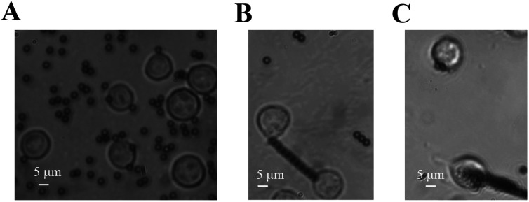
Biomolecules and organelles usually undergo changes to their structure or form as a result of mechanical stretching or stimulation. It is critical to be able to observe these changes and responses, which trigger mechano-chemical coupling or signal transduction. Advanced techniques have been developed to observe structure and form during manipulation; however, these require sophisticated methods. We have developed a simple approach to observe fine structure after stretching without fluorophore labeling. DNAs or molecules on the cell surface were bound to magnetic microbeads, followed by stretching with a magnetic field. After fixing, staining, and drying, the samples were examined by scanning electron microscopy with no need to build a functional surface with complex processes. Straight DNAs were observed rather than random-walk-like loose polymers. In our cellular experiment, the magnetic beads were bound to a Jurkat cell and formed a rosette which was later stuck to the substrate. A 41.3 μm filament on the base of a filopodium was pulled out via integrin from a cell. Therefore, our method can reveal long structures up to hundreds of micrometers at nanometer resolution after stretching or twisting. Our approach could have wide applications in structure-function studies of biomolecules, and in mechanobiology and cell biology when diffraction cannot used.
This journal is © The Royal Society of Chemistry.
Conflict of interest statement
There are no conflicts to declare.
Figures


Similar articles
-
Microfilaments in cellular and developmental processes.Science. 1971 Jan 15;171(3967):135-43. doi: 10.1126/science.171.3967.135. Science. 1971. PMID: 5538822
-
Pepstatin A: polymerization of an oligopeptide.Micron. 1994;25(2):189-217. doi: 10.1016/0968-4328(94)90042-6. Micron. 1994. PMID: 8055247 Review.
-
Twisting integrin receptors increases endothelin-1 gene expression in endothelial cells.Am J Physiol Cell Physiol. 2001 Jun;280(6):C1475-84. doi: 10.1152/ajpcell.2001.280.6.C1475. Am J Physiol Cell Physiol. 2001. PMID: 11350743
-
Visualization of actin filaments in keratocyte lamellipodia: negative staining compared with freeze-drying.J Struct Biol. 1994 Sep-Oct;113(2):135-41. doi: 10.1006/jsbi.1994.1043. J Struct Biol. 1994. PMID: 7718363
-
Stretch in Focus: 2D Inplane Cell Stretch Systems for Studies of Cardiac Mechano-Signaling.Front Bioeng Biotechnol. 2019 Mar 27;7:55. doi: 10.3389/fbioe.2019.00055. eCollection 2019. Front Bioeng Biotechnol. 2019. PMID: 30972334 Free PMC article. Review.
References
LinkOut - more resources
Full Text Sources