Review
. 2020 Mar 17;10(19):11095-11104.
doi: 10.1039/d0ra00950d. eCollection 2020 Mar 16.

Resonance Raman view of the active site architecture in bacterial DyP-type peroxidases

Affiliations
Review

Resonance Raman view of the active site architecture in bacterial DyP-type peroxidases

Célia M Silveira et al. RSC Adv. .

Abstract

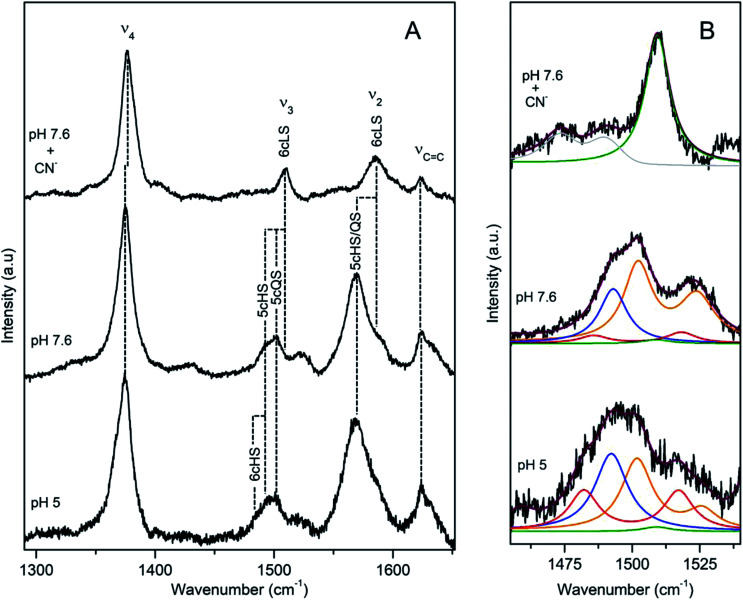
Dye decolorizing peroxidases (DyPs) are novel haem-containing peroxidases, which are structurally unrelated to classical peroxidases. They lack the highly conserved distal histidine that acts as an acid-base catalyst in the catalytic reaction of classical peroxidases, which implies distinct mechanistic properties. Despite the remarkable catalytic properties and recognized potential for biotechnology applications, the knowledge of DyP's structural features in solution, which govern the reactivity and catalysis, is lagging behind. Resonance Raman (RR) spectroscopy can reveal fine details of the active site structure in hemoproteins, reporting on the oxidation and spin state and coordination of the haem cofactor. We provide an overview of the haem binding pocket architecture of the enzymes from A, B and C DyP subfamilies, in the light of those established for classical peroxidases and search for subfamily specific features among DyPs. RR demonstrates that multiple spin populations typically co-exist in DyPs, like in the case of classical peroxidases. The haem spin/coordination state is strongly pH dependent and correlates well with the respective catalytic properties of DyPs. Unlike in the case of classical peroxidases, a surprisingly high abundance of catalytically incompetent low spin population is observed in several DyPs, and tentatively related to the alternative physiological function of these enzymes. The molecular details of active sites of DyPs, elucidated by RR spectroscopy, can furthermore guide approaches for biotechnological exploitation of these promising biocatalysts.


Conflict of interest statement

There are no conflicts to declare.

Figures

Fig. 1
Fig. 1. Detail of the haem binding pocket of Cbo, Tfu, Pp and Vc DyPs indicating the proximal His and the conserved distal residues Asp/Glu, Asn and Arg.
Fig. 2
Fig. 2. RR spectra of ferric BsDyP. (A) High frequency region for the resting state enzyme at pH 7.6 in the presence/absence of imidazole and at pH 5. (B) Component analysis of ν338 region; the component spectra represent the 6cLS (green) ν3 1506 cm−1, 6cHS (red) ν3 1481 cm−1 and ν38 1516 cm−1, 5cHS (blue) ν3 1488 cm−1 populations, overall fit (purple) and non-assigned bands (gray). Spectra were measured with 413 nm excitation.
Fig. 3
Fig. 3. RR spectra of ferric CboDyP. High frequency region for the resting state enzyme at pH 7.5 in the presence/absence of CN and at pHopt (pH 5). Spectra were measured with 413 nm excitation; experimental details can be found in ESI.
Fig. 4
Fig. 4. RR spectra of ferric TfuDyP. High frequency region of TfuDyP in the resting state at pH 7.5 in the presence/absence of CN and at pHopt (pH 3.5). Inset: component analysis of ν338 region; the component spectra represent the 6cHS (red) ν3 1481 cm−1 and ν38 1516 cm−1, 5cHS (blue) ν3 1493 cm−1 populations and the overall fit (purple). Spectra were measured with 413 nm excitation; experimental details can be found in ESI.
Fig. 5
Fig. 5. RR spectra of ferric PpDyP. (A) High frequency region for the resting state enzyme at pH 7.6 in the presence/absence of CN and at pHopt. (B) Component analysis of ν338 region; the component spectra represent the 6cLS (green) ν3 1509 cm−1, 6cHS (red) ν3 1483 cm−1 and ν38 1516 cm−1, 5cHS (blue) ν3 1493 cm−1, 5cQS (orange) ν3 1502 cm−1 and ν38 1525 cm−1 populations, overall fit (purple) and non-assigned bands (gray). Spectra were measured with 413 nm excitation.
Fig. 6
Fig. 6. RR spectra of VcDyP in the low-frequency (left) and high-frequency (right) regions. Spectra of ferric (a and d), ferrous (b and e), and ferrous CO-bound (c and f) forms are measured with 413.1 nm excitation wavelength for ferric and CO-bound forms and 441.6 nm for the ferrous form at pH 8. Reprinted with permission from (T. Uchida, et al., Biochemistry, 2015, 54, 6610–6621). Copyright (2020) American Chemical Society.
Fig. 7
Fig. 7. RR spectra of ferric DrDyP. High frequency region for the resting enzyme at pH 7.5 in the presence/absence of CN and at pH 4. The component spectra represent the 6cLS (green) and 6cHS (red) populations, overall fit (purple) and non-assigned bands (gray). Spectra were measured with 413 nm excitation; experimental details can be found in ESI.

References

    1. Sugano Y. Cell. Mol. Life Sci. 2009;66:1387–1403. doi: 10.1007/s00018-008-8651-8. - DOI - PMC - PubMed
    1. Yoshida T. Sugano Y. Arch. Biochem. Biophys. 2015;574:49–55. doi: 10.1016/j.abb.2015.01.022. - DOI - PubMed
    1. Singh R. Eltis L. D. Arch. Biochem. Biophys. 2015;574:56–65. doi: 10.1016/j.abb.2015.01.014. - DOI - PubMed
    1. Linde D. Pogni R. Cañellas M. Lucas F. Guallar V. Baratto M. C. Sinicropi A. Sáez-Jiménez V. Coscolín C. Romero A. Medrano F. J. Ruiz-Dueñas F. J. Martínez A. T. Biochem. J. 2015;466:253–262. doi: 10.1042/BJ20141211. - DOI - PMC - PubMed
    1. Colpa D. I. Fraaije M. W. van Bloois E. J. Ind. Microbiol. Biotechnol. 2014;41:1–7. doi: 10.1007/s10295-013-1371-6. - DOI - PubMed