Review
. 2020 Apr 20;10(26):15430-15460.
doi: 10.1039/d0ra00762e. eCollection 2020 Apr 16.

Applications of nano-materials in diverse dentistry regimes

Affiliations
Review

Applications of nano-materials in diverse dentistry regimes

Loke Kok Foong et al. RSC Adv. .

Abstract

Research and development in the applied sciences at the atomic or molecular level is the order of the day under the domain of nanotechnology or nano-science with enormous influence on nearly all areas of human health and activities comprising diverse medical fields such as pharmacological studies, clinical diagnoses, and supplementary immune system. The field of nano-dentistry has emerged due to the assorted dental applications of nano-technology. This review provides a brief introduction to the general nanotechnology field and a comprehensive overview of the synthesis features and dental uses of nano-materials including current innovations and future expectations with general comments on the latest advancements in the mechanisms and the most significant toxicological dimensions.


Conflict of interest statement

There are no conflicts to declare.

Figures

Fig. 1
Fig. 1. Representative structures of some nano-materials: (a) nanorings, (b) nanopellets, (c) nanorods, (d) nanosprings, (e) nanonails, (f) nanoflower.
Scheme 1
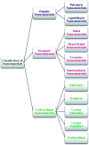
Scheme 1. General classification of nano-materials.
Scheme 2
Scheme 2. Conventional synthetic techniques for nano-materials.
Fig. 2
Fig. 2. Schematic diagram of the rechargeable nanoparticles of the amorphous calcium phosphate (NACP) sealant approach to deal with enamel demineralization around the dental sealants: In (A), the recharge cycle diagram illustrates the re-release from the exhausted and recharged NACP sealants. Three recharge/re-release cycles were performed and each re-release was measured for 14 days. The ion re-release increased on increasing the NACP filler level. In (B), the TEM image of NACP from the spray-drying technique, having sizes of about 100–300 nm.
Fig. 3
Fig. 3. Schematic representation of the anodization setup and the development of TiO2 NTs as well as PVD decoration of Ag2O NPs on the nanotubular layer.
Fig. 4
Fig. 4. Schematic diagram of PLGA (Ag–Fe3O4)-coated on dental implants.
Fig. 5
Fig. 5. Antibacterial and bioactive properties of stabilized silver on titanium with a nanostructured surface for dental implants.
Fig. 6
Fig. 6. Schematic illustration of the fabrication process of poly(lactic-co-glycolic acid)/Ag/ZnO nanorods composite coating.
Fig. 7
Fig. 7. Phytosynthesis of gold nanoparticles and evaluation of its osteoinductive potential for application in the implant dentistry.
Fig. 8
Fig. 8. Schematic illustration of the preparation of Ag NPs and the hybrid dental resin.
Fig. 9
Fig. 9. The preparation route of cellulose nanocrystal/zinc oxide (CNC/ZnO) nanohybrids.
Fig. 10
Fig. 10. Bottom-up controlled self-assembly of drug-silica mesoporous nanoparticles enables the long-term release of the antimicrobial agent at the site of recurrent caries: the restoration of the tooth interface. Utilizing a surfactant-like antimicrobial agent, mesoporous silica nanoparticles are inherently maximally loaded with the drug and exhibit extended controlled release. When incorporated into a dental restorative resin adhesive, these mesoporous silica nanoparticles release drug within the restoration-tooth marginal interface, killing caries-causing bacteria, thus extending the service life of the restoration through recurrent caries prevention.
Fig. 11
Fig. 11. Fabrication and in vivo evaluation of the hydroxyapatite/carbon nanotube electrospun fibers for biomedical/dental applications.
Fig. 12
Fig. 12. Technology for steering and delivering the drug-laden nanoparticles to the tooth pulp is described. This technology exploits naturally occurring dentinal tubules that extend from the dentin to the pulp and magnetic forces to actively steer the iron nanoparticles deep into the tooth structure. This technology was tested on rat molar teeth and in freshly extracted human teeth, and can be used to deliver drug-laden nanoparticles to the pulp or to enhance the bond strength of commercially available resin adhesives to the tooth dentin.

References

    1. Wu J. Wang X. Khatami M. Nejad M. S. Salari S. Zhu P. G. N. IET Nanobiotechnol. 2016;10:237. doi: 10.1049/iet-nbt.2015.0078. - DOI - PMC - PubMed
    1. Granjeiro J. M., Cruz R., Leite P. E., Gemini-Piperni S., Boldrini L. C. and Ribeiro A. R., Tin Oxide Materials ,Elsevier, 2020, p. 133
    1. Han J. Xiong L. Jiang X. Yuan X. Zhao Y. Yang D. Prog. Polym. Sci. 2019;91:1. doi: 10.1016/j.progpolymsci.2019.02.006. - DOI
    1. Pramanik S. and Sundar Das D., Two-Dimensional Nanostructures for Biomedical Technology, Elsevier, 2020, p. 281
    1. Liu P. Williams J. R. Cha J. J. Nat. Rev. Mater. 2019;4:479. doi: 10.1038/s41578-019-0113-4. - DOI