. 2021 Jun 2;4(6):1863-1891.
doi: 10.1016/j.matt.2021.03.013.

The Cell as Matter: Connecting Molecular Biology to Cellular Functions

Affiliations

The Cell as Matter: Connecting Molecular Biology to Cellular Functions

Yiwei Li et al. Matter. .

Abstract

Viewing cell as matter to understand the intracellular biomolecular processes and multicellular tissue behavior represents an emerging research area at the interface of physics and biology. Cellular material displays various physical and mechanical properties, which can strongly affect both intracellular and multicellular biological events. This review provides a summary of how cells, as matter, connect molecular biology to cellular and multicellular scale functions. As an impact in molecular biology, we review recent progresses in utilizing cellular material properties to direct cell fate decisions in the communities of immune cells, neurons, stem cells, and cancer cells. Finally, we provide an outlook on how to integrate cellular material properties in developing biophysical methods for engineered living systems, regenerative medicine, and disease treatments.


Conflict of interest statement

DECLARATION OF INTERESTS: The authors declare no competing interests.

Figures

Fig. 1.
Fig. 1.. Cells as a soft material exhibiting unique mechanical properties.
(A) Schematic illustration of cell compositions as passive matter or active matter. Reprinted with permission from Guo et al. Copyright 2014, Cell Press. (B) A diagram showing cytoplasmic mechanics as a function of frequency. Reprinted with permission from Gupta et al. Copyright 2017, Elsevier Publishing Company. (C) A diagram showing cytoplasmic dynamics, in terms of Mean Squared Displacement (MSD). Reprinted with permission from Gupta et al. Copyright 2017, Elsevier Publishing Company. (D) A diagram showing cytoplasmic forces, measured by Force Spectrum Microscopy (FSM). Reprinted with permission from Guo et al. Copyright 2014, Cell Press. (E) A state diagram summarizing different cytoplasmic mechanical behaviors, as either viscoelastic, poroelastic, pure viscous, or pure elastic materials, dependent of two dimensionless parameters, Deborah number (/a) and Péclet number (Va/D). Reprinted with permission from Hu et al. Copyright 2017, National Academy of Sciences.
Fig. 2.
Fig. 2.. Interplays between molecular crowding and cellular signaling transduction.
(A) Schematic illustration of the regulation of activation of YAP target genes by cellular/nuclear molecular crowding. Reprinted with permission from Cai et al. Copyright 2019, Nature Publishing Group. (B) Schematic illustration of the regulation of Wnt/β-catenin signaling via LRP6 signalosome by intracellular molecular crowding. Reprinted with permission from Li et al. Copyright 2021, Cell Press. (C) Schematic illustration of the regulation of intracellular molecular crowding by mTOR signaling via tuning intracellular ribosome concentration.
Fig. 3.
Fig. 3.. Regulation of cellular phase separation of biomacromolecules in a cellular physical property dependent manner.
(A) A state diagram shows crowding dependent phase separation of YAP protein. Reprinted with permission from Cai et al. Copyright 2019, Nature Publishing Group. (B) A state diagram shows the concentration and oligomerization dependent phase separation of cellular biomacromolecules. Reprinted with permission from Bracha et al. Copyright 2018, Cell Press. (C) Schematic illustration of tension and tissue spreading dependent phase separation of proteins in an embryo. Reprinted with permission from Schwayer et al. Copyright 2019, Cell Press. (D) Schematic illustration shows that the phase separation of biomolecular liquid droplets also generate force by surface tension to regulate genome architecture in cell nucleus. Reprinted with permission from Shin et al. Copyright 2018, Cell Press.
Fig. 4.
Fig. 4.. Physical properties of cell nucleus as a mechanoregulator of gene expression.
(A) Schematic illustration of an isolated intact nucleus. Reprinted with permission from Shivashankar et al. Copyright 2011, Annual Reviews. (B) Schematic illustration of nuclear deformation as a mechanoregulator downstream of cytoplasm-to-nucleus signaling transduction. Reprinted with permission from Elosegui-Artola et al. Copyright 2017, Cell Press. (C) Schematic illustration of nuclear deformation as a mechanoregulator of chromatin accessibility and gene expression. Reprinted with permission from Miroshnikova et al. Copyright 2017, The Company of Biologists Ltd.
Fig. 5.
Fig. 5.. Cellular components, that support cellular mechanical integrity, serve as key mechanoregulators of DNA damage.
Reprinted with permission from Shivashankar et al. Copyright 2011, Annual Reviews.
Fig. 6.
Fig. 6.. Regulations of cellular immunological processes depend on cellular physical properties.
(A) Schematic illustration of force and tension dependent T cell targeting killing in T cell immunological synapse. Reprinted with permission from Basu et al. Copyright 2016, Cell Press. (B) Schematic illustration of force-dependent antibodies uptake by B cells in B cell-Antigen-Presenting Cell (APC) immunological synapse. Reprinted with permission from Tolar et al. Copyright 2017, Nature Publishing Group. (C) Schematic illustration of nuclear swelling as a mechanoregulator of activation upstream of inflammation. Reprinted with permission from Enyedi et al. Copyright 2016, Cell Press. (D) Schematic illustration of cellular elasticity changes of immune cells during the inflammation process induced by different inflammatory factors. Reprinted with permission from Bufi et al. Copyright 2015, Cell Press.
Fig. 7.
Fig. 7.. Physical regulation of neuron fate decision and cellular physical property evolution during neurological lineage commitment.
(A) Schematic illustration of physical regulation by varying stiffness of extracellular matrix on neuron cells self-renewing and differentiating. Reprinted with permission from Tsai et al. Copyright 2019, Wiley Publishing Company. (B) Plot of cell area against deformation shows the cellular physical property changes during neuron lineage commitment from iPSCs. Reprinted with permission from Urbanska et al. Copyright 2017, The Company of Biologists Ltd. (C) Schematic illustration of cellular regulatory components that are involved in mechanoregulation in neuron cells. Reprinted with permission from Tsai et al. Copyright 2019, Wiley Publishing Company.
Fig. 8.
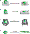
Fig. 8.. Various physical properties of cells on the regulation of fate decision of mesenchymal stem cells (MSCs) and embryonic stem cells (ESCs).
Reprinted with permission from Mosqueira et al. Copyright 2014, ACS publications. (A) Schematic illustration of volume reduction in MSCs or ESCs. (B) Schematic illustration of cell spreading in MSCs or ESCs. (C) Schematic illustration of cell stiffening in MSCs or ESCs. (D) Schematic illustration of cell elongation in MSCs or ESCs.
Fig. 9.
Fig. 9.. Different carcinogenic processes that transit cellular physical properties.
(A) Schematic illustration of epithelial-to-mesenchymal transitions (EMT) accompanying changes in cellular physical properties, such as adhesion, contractility, and spreading. Reprinted with permission from Dongre et al. Copyright 2019, Nature Publishing Group. (B) Schematic illustration of supercellular fluid flow driving changes in cellular physical properties, such as cell and nuclear volume, and cell stiffness. Reprinted with permission from Han et al. Copyright 2020, Nature Publishing Group. (C) Schematic illustration of jamming-unjamming transition accompanying changes in cellular physical properties, such as cellular aspect ratio, trajectory, perimeter, and area.

References

    1. Baluška F, Volkmann D & Barlow PW Cell bodies in a cage. Nature 428, 371–371 (2004). - PubMed
    1. Mazzarello P A unifying concept: the history of cell theory. Nature Cell Biology 1, E13–E15 (1999). - PubMed
    1. Zhou H-X, Rivas G & Minton AP Macromolecular crowding and confinement: biochemical, biophysical, and potential physiological consequences. Annu. Rev. Biophys 37, 375–397 (2008). - PMC - PubMed
    1. Alberts B et al. Molecular Biology of the Cell. third addition. (1994).
    1. Ho B, Baryshnikova A & Brown GW Unification of protein abundance datasets yields a quantitative Saccharomyces cerevisiae proteome. Cell systems 6, 192–205. e193 (2018). - PubMed

LinkOut - more resources