. 2022 Apr 12:13:834370.
doi: 10.3389/fmicb.2022.834370. eCollection 2022.

Linear Six-Carbon Sugar Alcohols Induce Lysis of Microcystis aeruginosa NIES-298 Cells

Affiliations

Linear Six-Carbon Sugar Alcohols Induce Lysis of Microcystis aeruginosa NIES-298 Cells

Jaejoon Jung et al. Front Microbiol. .

Abstract

Cyanobacterial blooms are a global concern due to their adverse effects on water quality and human health. Therefore, we examined the effects of various compounds on Microcystis aeruginosa growth. We found that Microcystis aeruginosa NIES-298 cells were lysed rapidly by linear six-carbon sugar alcohols including mannitol, galactitol, iditol, fucitol, and sorbitol, but not by other sugar alcohols. Microscopic observations revealed that mannitol treatment induced crumpled inner membrane, an increase in periplasmic space, uneven cell surface with outer membrane vesicles, disruption of membrane structures, release of intracellular matter including chlorophylls, and eventual cell lysis in strain NIES-298, which differed from the previously proposed cell death modes. Mannitol metabolism, antioxidant-mediated protection of mannitol-induced cell lysis by, and caspase-3 induction in strain NIES-298 were not observed, suggesting that mannitol may not cause organic matter accumulation, oxidative stress, and programmed cell death in M. aeruginosa. No significant transcriptional expression was induced in strain NIES-298 by mannitol treatment, indicating that cell lysis is not induced through transcriptional responses. Mannitol-induced cell lysis may be specific to strain NIES-298 and target a specific component of strain NIES-298. This study will provide a basis for controlling M. aeruginosa growth specifically by non-toxic substances.

Keywords: cell lysis; cyanobacterial bloom; mannitol; outer membrane vesicle; six-carbon sugar alcohol.


Conflict of interest statement

The authors declare that the research was conducted in the absence of any commercial or financial relationships that could be construed as a potential conflict of interest.

Figures

FIGURE 1
FIGURE 1
Effects of various organic compounds on the growth of Microcystis aeruginosa NIES-298. After adding organic compounds (50 μM), the cultures were incubated at 25°C under 40 μmol photons m–2⋅s–1 with a 12 h/12 h light/dark cycle. Data are indicated as mean values ± standard deviations. Statistical significances depending on organic compounds were calculated compared to the control group (no treatment) and their significant differences were indicated with different colors. **p < 0.01; ***p < 0.001.
FIGURE 2
FIGURE 2
The growth of Microcystis aeruginosa NIES-298 treated with sugar alcohols without any effect (A) and with inhibiting effect on the growth (B). After adding sugar alcohols (50 μM), the cultures were incubated at 25°C under 40 μmol photons m–2⋅s–1 with a 12 h/12 h light/dark cycle. Numbers in the parenthesis indicate the carbon numbers of sugar alcohols. Data are indicated as mean values ± standard deviations. Statistical significances depending on sugar alcohols were calculated compared to the control group (no treatment) and their significant differences were indicated with different colors. *p < 0.05; **p < 0.01; ***p < 0.001.
FIGURE 3
FIGURE 3
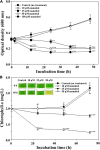
Optical cell density (A) and chlorophyll a (B) profiles of Microcystis aeruginosa NIES-298 treated with different concentrations of mannitol. Data are indicated as mean values ± standard deviations. Images showing the color changes of cell cultures after mannitol treatment are also displayed in (B). Statistical significances depending on the mannitol treatments were calculated compared to the control group (no treatment) and their significant differences were indicated with p-values < 0.05 (*), < 0.01 (**), or < 0.001 (***).
FIGURE 4
FIGURE 4
Time course caspase-3 (-like) activity of Microcystis aeruginosa NIES-298 cells treated with mannitol and H2O2 (as a positive control agent). The caspase activities were expressed as arbitrary unit (AU). Data are indicated as mean values ± standard deviations. Statistical significances depending on the treatments were calculated compared to the control group (no treatment) and their significant differences were indicated with p-values < 0.001 (***).
FIGURE 5
FIGURE 5
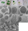
DICM (A–E), TEM (F–K), and SEM (L,M) images showing the morphological changes in Microcystis aeruginosa NIES-298 cells upon mannitol treatment (50 μM). Photographs (A,F,L) indicate intact cells without mannitol treatment and all mannitol-treated cells (B–E,G–K,M) were observed within 2 h (the SEM image in (M) was obtained at 2 h after mannitol treatment). After mannitol treatment, the proportion of damaged cells (F,K) increased over time. Open arrows, triangles, and closed arrows indicate the disruption of cell walls, outer membrane vesicles, and intracellular matter released from cells, respectively. Open arrows in (M) indicate pores formed on cell walls after mannitol treatment.
FIGURE 6
FIGURE 6
Concentration profiles of intracellular organic matter (A) and chlorophyll a (B) released from Microcystis aeruginosa NIES-298 cells treated with mannitol. After mannitol treatment, cell cultures were filtered using a 0.45-μm syringe filter and the concentrations of organic matter (254 nm) and chlorophyll a in the filtrates were measured. Data are indicated as mean values ± standard deviations. Statistical significances depending on the mannitol treatments were calculated compared to the control group (no treatment) and their significant differences were indicated with p-values < 0.01 (**) or < 0.001 (***).

References

    1. Alexova R., Fujii M., Birch D., Cheng J., Waite T. D., Ferrari B. C., et al. (2011). Iron uptake and toxin synthesis in the bloom-forming Microcystis aeruginosa under iron limitation. Environ. Microbiol. 13 1064–1077. 10.1111/j.1462-2920.2010.02412.x - DOI - PubMed
    1. Al-Tebrineh J., Merrick C., Ryan D., Humpage A., Bowling L., Neilan B. A. (2012). Community composition, toxigenicity, and environmental conditions during a cyanobacterial bloom occurring along 1,100 kilometers of the Murray River. Appl. Environ. Microbiol. 78 263–272. 10.1128/AEM.05587-11 - DOI - PMC - PubMed
    1. Berman-Frank I., Bidle K. D., Haramaty L., Falkowski P. G. (2004). The demise of the marine cyanobacterium, Trichodesmium spp., via an autocatalyzed cell death pathway. Limnol. Oceanogr. 49 997–1005. 10.4319/lo.2004.49.4.0997 - DOI
    1. Bhattacharjee S., Mishra A. K. (2020). The tale of caspase homologues and their evolutionary outlook: deciphering programmed cell death in cyanobacteria. J. Exp. Bot. 71 4639–4657. 10.1093/jxb/eraa213 - DOI - PMC - PubMed
    1. Biller S. J., Schubotz F., Roggensack S. E., Thompson A. W., Summons R. E., Chisholm S. W. (2014). Bacterial vesicles in marine ecosystems. Science 343 183–186. 10.1126/science.1243457 - DOI - PubMed