

Placental Tissues as Biomaterials in Regenerative Medicine
- PMID: 35496035
- PMCID: PMC9050314
- DOI: 10.1155/2022/6751456
Placental Tissues as Biomaterials in Regenerative Medicine
Abstract
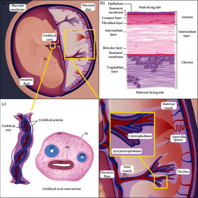
Placental tissues encompass all the tissues which support fetal development, including the placenta, placental membrane, umbilical cord, and amniotic fluid. Since the 1990s there has been renewed interest in the use of these tissues as a raw material for regenerative medicine applications. Placental tissues have been extensively studied for their potential contribution to tissue repair applications. Studies have attributed their efficacy in augmenting the healing process to the extracellular matrix scaffolds rich in collagens, glycosaminoglycans, and proteoglycans, as well as the presence of cytokines within the tissues that have been shown to stimulate re-epithelialization, promote angiogenesis, and aid in the reduction of inflammation and scarring. The compositions and properties of all birth tissues give them the potential to be valuable biomaterials for the development of new regenerative therapies. Herein, the development and compositions of each of these tissues are reviewed, with focus on the structural and signaling components that are relevant to medical applications. This review also explores current configurations and recent innovations in the use of placental tissues as biomaterials in regenerative medicine.
Copyright © 2022 Annelise Roy et al.
Conflict of interest statement
All authors declare that they are employees of StimLabs, LLC.
Figures
References
-
- Gupta S., Gupta R. Placental Tissues- From Reproductive to Regenerative Biology. Int. J. Sci. Res. IJSR . 2014;3:607–612.
Publication types
MeSH terms
Substances
LinkOut - more resources
Full Text Sources
Other Literature Sources
