. 2022 Apr 12:16:876204.
doi: 10.3389/fnhum.2022.876204. eCollection 2022.

Responsive Neurostimulation Targeting the Anterior, Centromedian and Pulvinar Thalamic Nuclei and the Detection of Electrographic Seizures in Pediatric and Young Adult Patients

Affiliations

Responsive Neurostimulation Targeting the Anterior, Centromedian and Pulvinar Thalamic Nuclei and the Detection of Electrographic Seizures in Pediatric and Young Adult Patients

Cameron P Beaudreault et al. Front Hum Neurosci. .

Abstract

Background: Responsive neurostimulation (RNS System) has been utilized as a treatment for intractable epilepsy. The RNS System delivers stimulation in response to detected abnormal activity, via leads covering the seizure foci, in response to detections of predefined epileptiform activity with the goal of decreasing seizure frequency and severity. While thalamic leads are often implanted in combination with cortical strip leads, implantation and stimulation with bilateral thalamic leads alone is less common, and the ability to detect electrographic seizures using RNS System thalamic leads is uncertain.

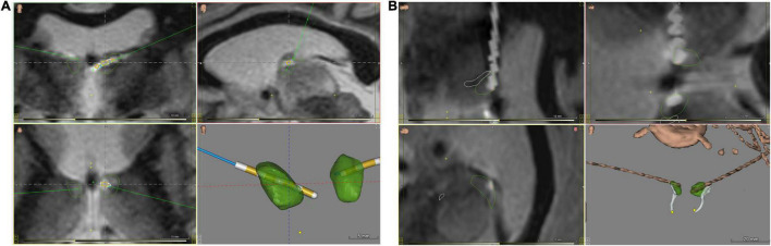
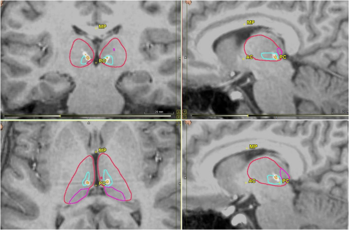
Objective: The present study retrospectively evaluated fourteen patients with RNS System depth leads implanted in the thalamus, with or without concomitant implantation of cortical strip leads, to determine the ability to detect electrographic seizures in the thalamus. Detailed patient presentations and lead trajectories were reviewed alongside electroencephalographic (ECoG) analyses.

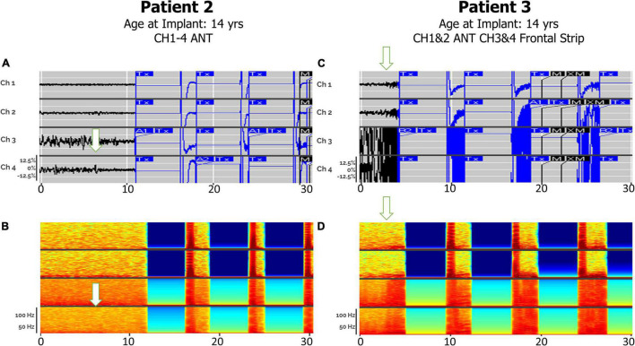
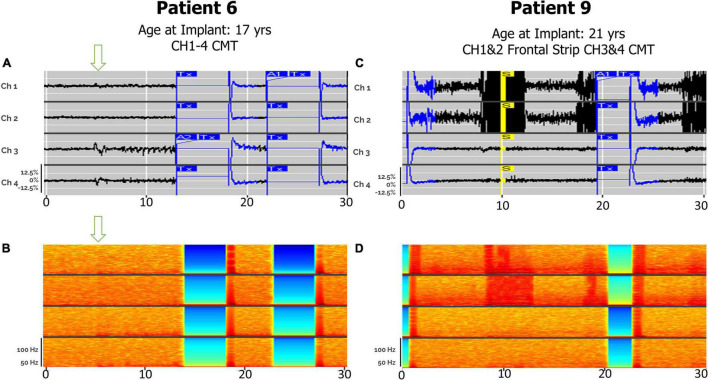
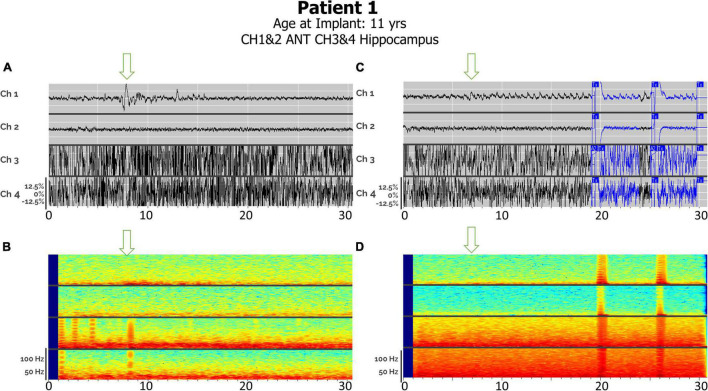
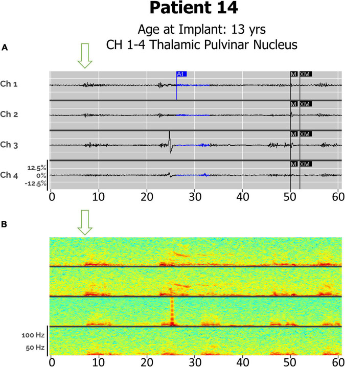
Results: Anterior nucleus thalamic (ANT) leads, whether bilateral or unilateral and combined with a cortical strip lead, successfully detected and terminated epileptiform activity, as demonstrated by Cases 2 and 3. Similarly, bilateral centromedian thalamic (CMT) leads or a combination of one centromedian thalamic alongside a cortical strip lead also demonstrated the ability to detect electrographic seizures as seen in Cases 6 and 9. Bilateral pulvinar leads likewise produced reliable seizure detection in Patient 14. Detections of electrographic seizures in thalamic nuclei did not appear to be affected by whether the patient was pediatric or adult at the time of RNS System implantation. Sole thalamic leads paralleled the combination of thalamic and cortical strip leads in terms of preventing the propagation of electrographic seizures.

Conclusion: Thalamic nuclei present a promising target for detection and stimulation via the RNS System for seizures with multifocal or generalized onsets. These areas provide a modifiable, reversible therapeutic option for patients who are not candidates for surgical resection or ablation.

Keywords: RNS; anterior thalamic nucleus; centromedian thalamic nucleus; epilepsy surgery; intractable epilepsy; pulvinar; responsive neurostimulation; thalamic stimulation.


Conflict of interest statement

SW and PM reports honoraria from LivaNova, Eisai, UCB, Sunovion, Greenwich Pharmaceuticals, and participation as an investigator in clinical trials for Zogenix, GW Pharma, NeuroPace, Neurelis, UCB, Eisai. CM reports speaker fees from LivaNova, and participation as an investigator in clinical trial for NeuroPace. The remaining authors declare that the research was conducted in the absence of any commercial or financial relationships that could be construed as a potential conflict of interest. The handling editor GW declared a past co-authorship with the author(s) SW and PM.

Figures

FIGURE 1
FIGURE 1
ANT nuclei images with brain atlas overlay of postoperative CT/MRI merge, visualizing implanted RNS depth electrodes. (A) Coronal (upper left and lower left) and sagittal (upper right) views, and 3D-rendered image of implanted ANT nuclei without background brain. (B) Close-up views of implanted leads from rotated sagittal (upper and lower left) and oblique (upper right) views, along with an oblique view 3D-rendered image of implanted ANT nuclei without background brain. ANT nuclei marked in green, mammillothalamic tracts in white, electrodes in brown. Images generated with WayPoint Navigator version 4.6.6.
FIGURE 2
FIGURE 2
Postoperative CMT nuclei images in coronal (upper left), axial (bottom left) and sagittal views (upper and lower right) with RNS electrodes visualized. CMT is outlined in light blue, thalamus in red, pulvinar in pink, and electrode in white with each contact in orange. Images generated using WayPoint Navigator version 4.6.6.
FIGURE 3
FIGURE 3
Electrographic seizures detected in ANT (Anterior Thalamic Nucleus), with and without cortical strip leads, recorded by the RNS system. Arrows denote seizure onsets. (A) An example of a clinical seizure in Patient 2, stored in the NeuroPace Patient Data Management System (PDMS) over a 30-s window with bilateral ANT leads, left channels (Ch 1. L-ANT1–L-ANT2; Ch 2. L-ANT3–L-ANT4) and right channels (Ch 3. R-ANT1–R-ANT2; Ch 4. R-ANT3–R-ANT4). (B) Spectrogram of identical epoch. (C) An example of an electroclinical generalized tonic-clonic seizure in Patient 3 stored in PDMS over a 30-s window, with a right ANT depth lead and a right anterior prefrontal cortical strip (Anc) lead. Ch 1. R-ANT1–R-ANT2; Ch 2. R-ANT3–R-ANT4; Ch 3. R-Anc1–R-Anc2; Ch 4. R-Anc3–R-Anc4. (D) Spectrogram of identical epoch. Tx, therapy; M, magnet swipe; A1, Pattern A, 1st detector; B2, Pattern B, 2nd detector; M, magnet; XM, magnet removed.
FIGURE 4
FIGURE 4
Electroclinical seizures detected in CMT, with and without cortical strip leads, recorded by the RNS system over a 30-s window. Arrows denote seizure onsets. (A) An example of an electroclinical seizure consisting of myoclonic jerks in Patient 6, captured with bilateral CMT leads over a 30-s window. Ch 1. R-CMT1–R-CMT2; Ch 2. R-CMT3–R-CMT4; Ch 3. L-CMT1–L-CMT2; Ch 4. L-CMT3–L-CMT4. (B) Spectrogram of identical epoch. (C) An example of an electroclinical seizure in Patient 9 that was ongoing at the time of capture, detected with left frontal cortical strip (Fnt) + right CMT depth leads over a 30-s window with saturation in the cortex and subsequent spread to CMT thalamic leads. Ch 1. L-Fnt1–L-Fnt2; Ch 2. L-Fnt3–L-Fnt4; Ch 3. R-CMT1–R-CMT2; Ch 4. R-CMT3–R-CMT4. (D) Spectrogram of identical epoch. S, Saturation; A2, Pattern A, 2nd detector.
FIGURE 5
FIGURE 5
(A) Electroclinical detection of a focal sensory seizure over a 30-s window in Patient #1, with arrows denoting seizure onsets in thalamic lead after seizure onset in hippocampus has already started. Ch 1. R-ANT1–R-ANT2; Ch 2. R-ANT3–R-ANT4; Ch 3. L-Hip–1–L-Hip2; Ch 4. L-Hip3–LHip4). (B) Spectrogram of same epoch. (C) Electroclinical seizure detection and treatment of generalized tonic clonic seizure pattern over a 30-s window with L-Hippocampus onset already started prior to this clip but note thalamic seizure activity onset by the arrow. (D) Spectrogram of same epoch. Tx, therapy; T–c, Charge insufficient.
FIGURE 6
FIGURE 6
(A) Electroclinical seizure detections in Patient 14 captured with bilateral pulvinar (Pulv) leads over an 60-s window, demonstrating both a clinical generalized seizures (Drop), with in-phase waveforms, and focal seizures (left arm fencing posture), with out-of-phase waveforms. Arrows denote seizure onsets. Ch1. L-Pulv1–L-Pulv2; Ch 2. L-Pulv3–LPulv4; Ch 3. R-Pulv1–R-Pulv2; Ch 4. R-Pulv3–R-Pulv4. (B) Spectrogram of same epoch.

References

    1. Bergey G. K., Morrell M. J., Mizrahi E. M., Goldman A., King-Stephens D., Nair D., et al. (2015). Long-term treatment with responsive brain stimulation in adults with refractory partial seizures. Neurology. 84 810–817. - PMC - PubMed
    1. Burdette D., Mirro E. A., Lawrence M., Patra S. E. (2021). Brain-responsive corticothalamic stimulation in the pulvinar nucleus for the treatment of regional neocortical epilepsy: a case series. Epilepsia Open. 6 611–617. 10.1002/epi4.12524 - DOI - PMC - PubMed
    1. Burdette D. E., Haykal M. A., Jarosiewicz B., Fabris R. R., Heredia G., Elisevich K., et al. (2020). Brain-responsive corticothalamic stimulation in the centromedian nucleus for the treatment of regional neocortical epilepsy. Epilepsy Behav. 112:107354. 10.1016/j.yebeh.2020.107354 - DOI - PubMed
    1. Elder C., Friedman D., Devinsky O., Doyle W., Dugan P. (2019). Responsive neurostimulation targeting the anterior nucleus of the thalamus in 3 patients with treatment-resistant multifocal epilepsy. Epilepsia Open. 4 187–192. 10.1002/epi4.12300 - DOI - PMC - PubMed
    1. Engel J., Jr., McDermott M. P., Wiebe S., Langfitt J. T., Stern J. M., Dewar S., et al. (2012). Early surgical therapy for drug-resistant temporal lobe epilepsy: a randomized trial. JAMA. 307 922–930. 10.1001/jama.2012.220 - DOI - PMC - PubMed

LinkOut - more resources