

Icariin controlled release on a silk fibroin/mesoporous bioactive glass nanoparticles scaffold for promoting stem cell osteogenic differentiation
- PMID: 35496600
- PMCID: PMC9050898
- DOI: 10.1039/d0ra00637h
Icariin controlled release on a silk fibroin/mesoporous bioactive glass nanoparticles scaffold for promoting stem cell osteogenic differentiation
Abstract
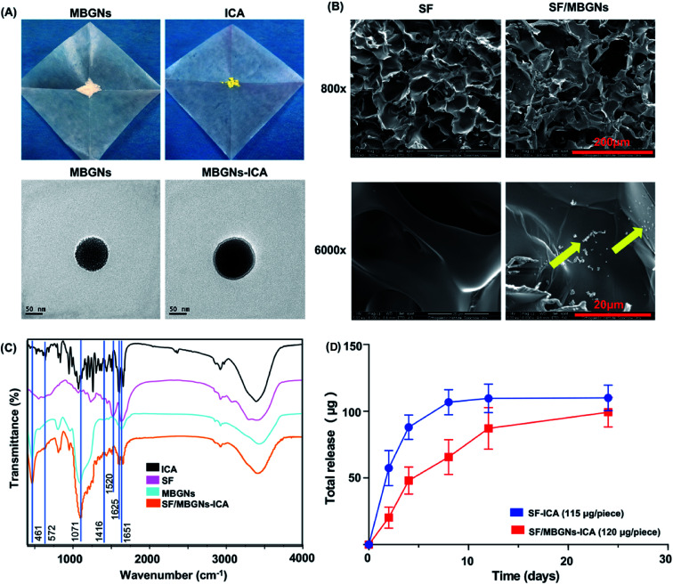
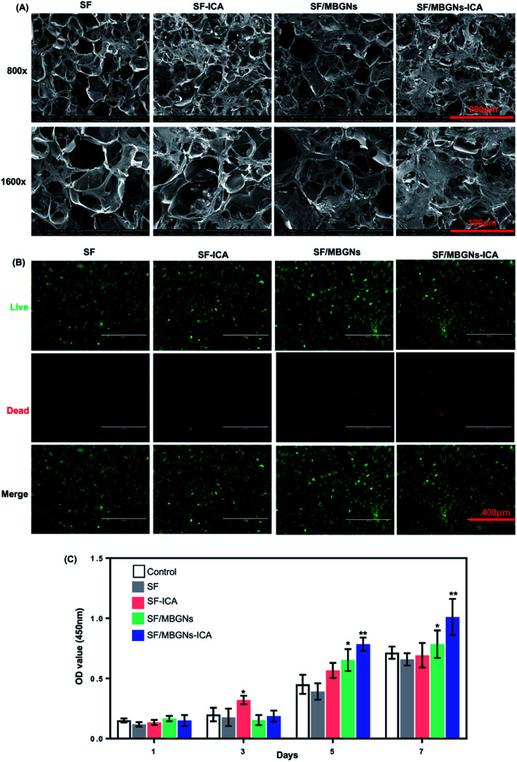
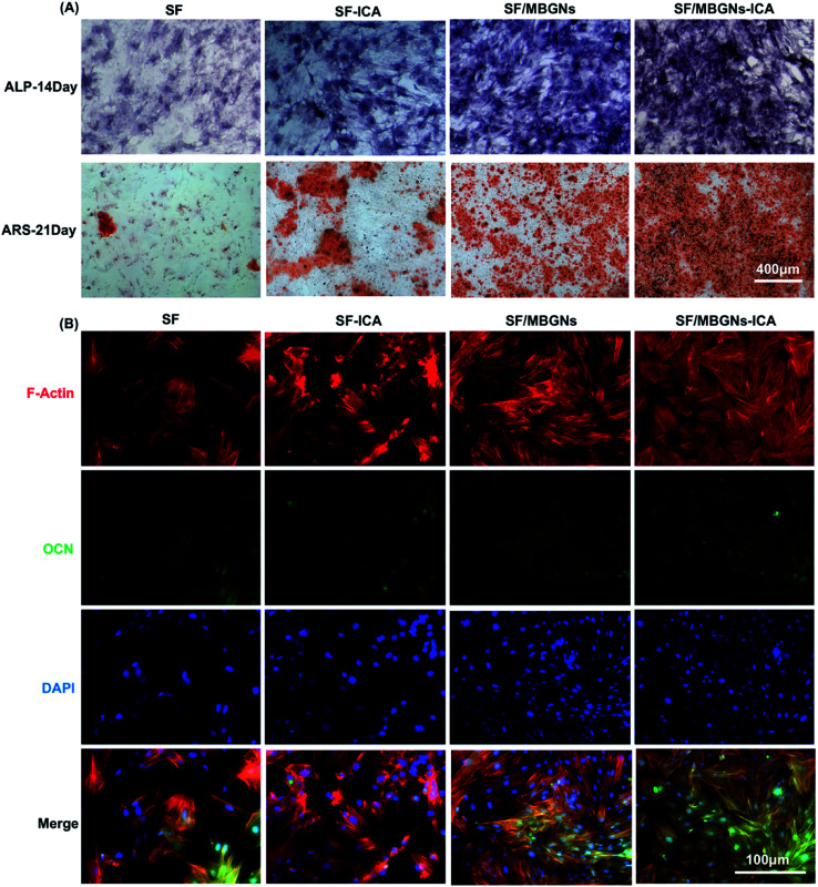
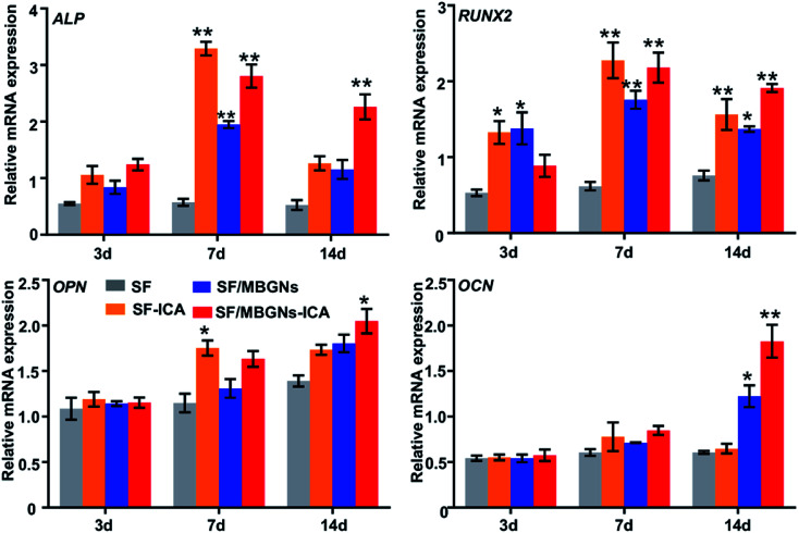
The treatment of bone defects caused by various reasons is still a major problem in orthopedic clinical work. Many studies on osteogenic implant materials have used various biologically active factors such as osteogenic inducers, but these biologically active factors have various side effects. Therefore, in this study, silk fibroin (SF) was used as a scaffold material, mesoporous bioactive glass nanoparticles (MBGNs) as a sustained release carrier, and the traditional Chinese drug icariin (ICA) was loaded to promote bone formation. The experiments in this study have proven that SF/MBGNs-ICA scaffolds can successfully load and release ICA for a long time, and the sustained-release ICA can promote the proliferation and differentiation of BMSCs for a long time. This controlled-release ICA organic/inorganic two-component scaffold material is expected to become a new bone grafting solution.
This journal is © The Royal Society of Chemistry.
Conflict of interest statement
The authors declare no conflicts of interest.
Figures
Similar articles
-
Mesoporous Hydroxyapatite Nanoparticles Mediate the Release and Bioactivity of BMP-2 for Enhanced Bone Regeneration.ACS Biomater Sci Eng. 2020 Apr 13;6(4):2323-2335. doi: 10.1021/acsbiomaterials.9b01954. Epub 2020 Mar 27. ACS Biomater Sci Eng. 2020. PMID: 33455303
-
Osteogenic properties of manganese-doped mesoporous bioactive glass nanoparticles.J Biomed Mater Res A. 2020 Sep;108(9):1806-1815. doi: 10.1002/jbm.a.36945. Epub 2020 Apr 21. J Biomed Mater Res A. 2020. PMID: 32276292
-
Fabrication of hierarchically porous silk fibroin-bioactive glass composite scaffold via indirect 3D printing: Effect of particle size on physico-mechanical properties and in vitro cellular behavior.Mater Sci Eng C Mater Biol Appl. 2019 Oct;103:109688. doi: 10.1016/j.msec.2019.04.067. Epub 2019 Apr 22. Mater Sci Eng C Mater Biol Appl. 2019. PMID: 31349405
-
Periosteum and development of the tissue-engineered periosteum for guided bone regeneration.J Orthop Translat. 2022 Feb 16;33:41-54. doi: 10.1016/j.jot.2022.01.002. eCollection 2022 Mar. J Orthop Translat. 2022. PMID: 35228996 Free PMC article. Review.
-
Silk fibroin scaffolds: A promising candidate for bone regeneration.Front Bioeng Biotechnol. 2022 Nov 25;10:1054379. doi: 10.3389/fbioe.2022.1054379. eCollection 2022. Front Bioeng Biotechnol. 2022. PMID: 36507269 Free PMC article. Review.
Cited by
-
Engineering mesoporous bioactive glasses for emerging stimuli-responsive drug delivery and theranostic applications.Bioact Mater. 2024 Jan 12;34:436-462. doi: 10.1016/j.bioactmat.2024.01.001. eCollection 2024 Apr. Bioact Mater. 2024. PMID: 38282967 Free PMC article. Review.
-
[Research progress on biocomposites based on bioactive glass].Sheng Wu Yi Xue Gong Cheng Xue Za Zhi. 2023 Aug 25;40(4):805-811. doi: 10.7507/1001-5515.202202016. Sheng Wu Yi Xue Gong Cheng Xue Za Zhi. 2023. PMID: 37666773 Free PMC article. Review. Chinese.
-
A Review of Bioactive Glass/Natural Polymer Composites: State of the Art.Materials (Basel). 2020 Dec 6;13(23):5560. doi: 10.3390/ma13235560. Materials (Basel). 2020. PMID: 33291305 Free PMC article. Review.
-
Promoting osteogenesis and bone regeneration employing icariin-loaded nanoplatforms.J Biol Eng. 2024 Apr 22;18(1):29. doi: 10.1186/s13036-024-00425-4. J Biol Eng. 2024. PMID: 38649969 Free PMC article. Review.
-
The Role of Herbal Medicine in Modulating Bone Homeostasis.Curr Top Med Chem. 2024;24(7):634-643. doi: 10.2174/0115680266286931240201131724. Curr Top Med Chem. 2024. PMID: 38333981 Review.
References
-
- Li Z. Xie M. B. Li Y. Ma Y. Li J. S. Dai F. Y. J. Biomater. Tissue Eng. 2016;12:755–766. doi: 10.1166/jbt.2016.1510. - DOI
LinkOut - more resources
Full Text Sources
Miscellaneous