

Spontaneous hyphema from iris microhemangioma in Eisenmenger syndrome
- PMID: 35496761
- PMCID: PMC9046946
- DOI: 10.1016/j.ajoc.2022.101536
Spontaneous hyphema from iris microhemangioma in Eisenmenger syndrome
Abstract
Purpose: We describe a patient with Eisenmenger syndrome and spontaneous hyphema from iris microhemangioma, two rare entities with a plausible pathophysiological connection.
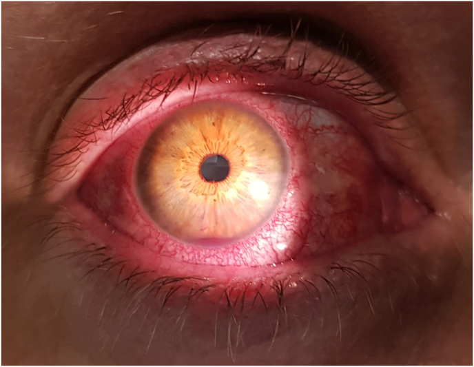
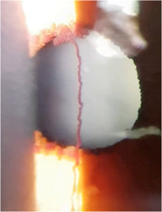
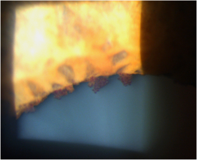
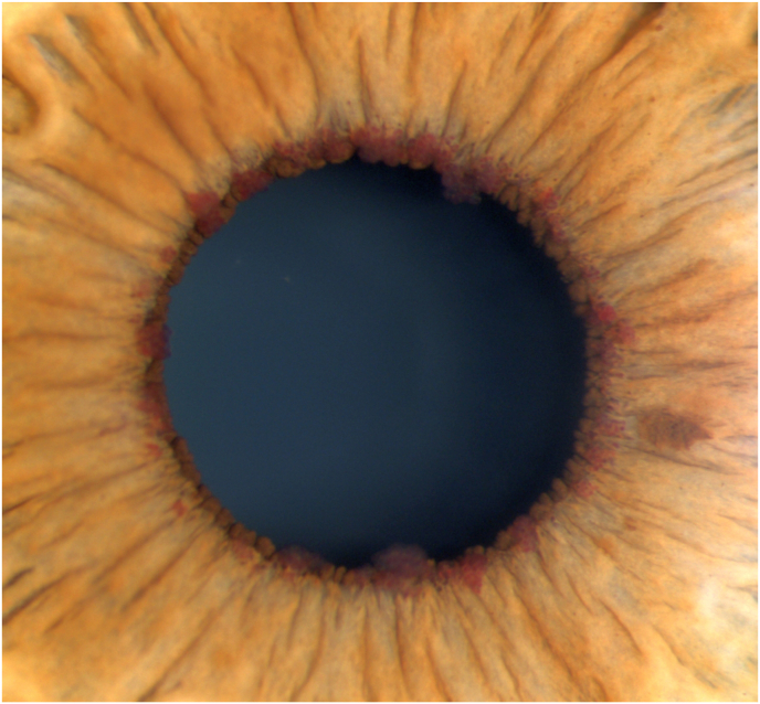
Observations: A 56-year-old Caucasian female with a background of cyanotic congenital heart disease complicated by Eisenmenger syndrome presented with non-traumatic hyphema and blurred vision. Multiple vascular tufts consistent with iris microhemangiomas were observed around the pupil margins bilaterally, with no iris or retinal neovascularization. In the affected eye, there was active bleeding from one lesion at 12 o'clock generating a macrohyphema. Additional findings included prominent episcleral injection and retinal venous tortuosity in both eyes. The active microhemorrhage and hyphema resolved with local medical management.
Conclusions and importance: Chronic hypoxemia and erythrocytosis are known to induce dilation of the retinal and episcleral blood vessels in Eisenmenger syndrome. Corresponding dilation of iris stromal vessels may contribute to the formation and prominence of iris microhemangiomas.
Keywords: Cobb's tuft; Eisenmenger syndrome; Iris microhemangioma; Iris vascular tuft; Spontaneous hyphema.
© 2022 Published by Elsevier Inc.
Figures
References
-
- Bakke E.F., Drolsum L. Iris microhaemangiomas and idiopathic juxtafoveolar retinal telangiectasis. Acta Ophthalmol Scand. 2006;84(6):818–822. - PubMed
-
- Nasir-Ahmad S., Cordina R., Liew G., McCluskey P., Celermajer D. The eye in CHD. Cardiol Young. 2018;28(8):981–985. - PubMed
-
- Cobb B., Shilling J.S., Chisholm I.H. Vascular tufts at the pupillary margin in myotonic dystrophy. Am J Ophthalmol. 1970;69(4):573–582. - PubMed
Publication types
LinkOut - more resources
Full Text Sources
