Case Reports
. 2022 Apr 10:26:101534.
doi: 10.1016/j.ajoc.2022.101534. eCollection 2022 Jun.

Ocular involvement in TEMPI syndrome

Affiliations
Case Reports

Ocular involvement in TEMPI syndrome

Jo-Hsuan Wu et al. Am J Ophthalmol Case Rep. .

Abstract

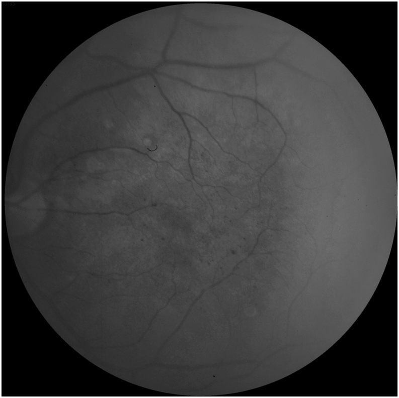
Purpose: We report the first case of ocular involvement in TEMPI syndrome, a rare disease characterized by telangiectasias, elevated erythropoietin with erythrocytosis, monoclonal gammopathy, perinephric fluid collections, and intra-pulmonary shunting.

Observations: A 64-year-old Caucasian man with history of TEMPI syndrome presented with subacute bilateral painless vision loss. Ocular examination showed chronic retinal ischemia with microvascular damage, which was likely associated with the chronic systemic hypoxemia, and spontaneous wax and wane of cystoid macular edema, presumedly related to the systemic bortezomib treatment.

Conclusions and importance: Our case demonstrates that pathologic retinal vascular changes could be seen in association with TEMPI syndrome and suggests that a comprehensive ophthalmological examination may be beneficial for these patients.

Keywords: Macular edema; Retinal ischemia; TEMPI syndrome.


Conflict of interest statement

No conflicting relationship exists for any authors.

Figures

Fig. 1
Fig. 1
Optical coherence tomography images at presentation (A, OD; B, OS), 1-week follow up (C, OD; D, OS), and 2-month follow up (E, OD; F, OS), showing spontaneous waxing and waning of cystoid macular edema.
Fig. 2
Fig. 2
Optical coherence tomography of the right eye showed focal inner retinal thinning, consistent with prior retinal ischemia (blue arrow head). (For interpretation of the references to colour in this figure legend, the reader is referred to the Web version of this article.)
Fig. 3
Fig. 3
Red free photograph of the left eye showed numerous parafoveal microaneurysms.
Fig. 4
Fig. 4
Fluorescein angiography of the right eye (A) showed microaneurysms and diffuse leakage, while the left eye (B) showed an enlarged foveal avascular zone, areas of retinal non-perfusion, vascular anastomoses, and diffuse leakage.

References

    1. Sykes D.B., Schroyens W., O'Connell C. The TEMPI syndrome — a novel multisystem disease. N Engl J Med. 2011;365(5):475–477. doi: 10.1056/NEJMc1106670. - DOI - PubMed
    1. Kwok M., Korde N., Landgren O. Bortezomib to treat the TEMPI syndrome. N Engl J Med. 2012;366(19):1843–1845. doi: 10.1056/NEJMc1202649. - DOI - PMC - PubMed
    1. Ryden A., Wei K., Rodriguez R., Mahrer T. Too much blood: a case of the newly described TEMPI syndrome. Chest. 2013/10/01/2013;144(4, Supplement):927A. doi: 10.1378/chest.1701121. - DOI
    1. Jasim S., Mahmud G., Bastani B., Fesler M. Subcutaneous bortezomib for treatment of TEMPI syndrome. Clin Lymphoma, Myeloma & Leukemia. Dec 2014;14(6):e221–e223. doi: 10.1016/j.clml.2014.07.004. - DOI - PubMed
    1. Sykes D.B., Schroyens W. Complete responses in the TEMPI syndrome after treatment with daratumumab. N Engl J Med. 2018;378(23):2240–2242. doi: 10.1056/NEJMc1804415. - DOI - PubMed

Publication types

LinkOut - more resources