

doi: 10.1016/j.xpro.2022.101342.
eCollection 2022 Jun 17.
Detailed protocol for germ-free Drosophila melanogaster colonization with Propionibacterium spp. biofilms
Affiliations
- PMID: 35496790
- PMCID: PMC9046622
- DOI: 10.1016/j.xpro.2022.101342
Item in Clipboard
Detailed protocol for germ-free Drosophila melanogaster colonization with Propionibacterium spp. biofilms
STAR Protoc.
.
Abstract
In this protocol, we describe a germ-free Drosophila melanogaster model to investigate anaerobic bacterial biofilms. We detail how to establish Propionibacterium spp. biofilms in the fruit fly's gut using an easy to carry out method. For complete details on the use and execution of this protocol, please refer to Bronnec and Alexeyev (2021) and Bronnec et al. (2022).
Keywords: Health Sciences; Microbiology; Microscopy; Model Organisms.
© 2022 The Authors.
Conflict of interest statement
The authors declare no competing interests.
Figures

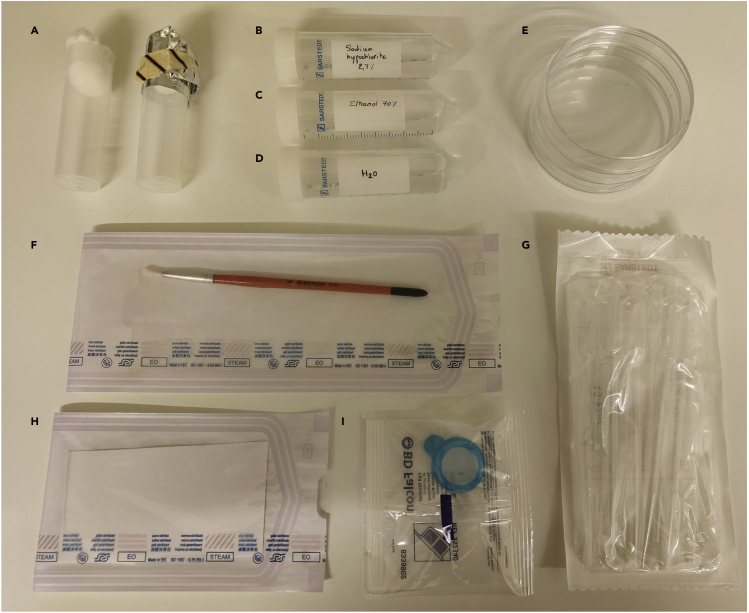
Disposable washing kit to generate germ-free Drosophila melanogaster (A) Fruit fly vials should be closed with a cotton plug and tightly wrap in aluminum foil for autoclaving. (B–D) Sodium hypochlorite and ethanol are diluted in sterile milli-Q water. All liquid solutions should be heated at 30°C before use. (E, G, and I) Plastic disposables purchased sterile. (F and H) Painting brush (washed in 70% ethanol) and blotting papers (disposable) are sterilized in autoclavable envelopes.

Germ-free Drosophila melanogaster line generation GF fruit flies are obtained from WT fruit flies after three generations (G1, G2 and G3) of washing and raising on sterile media with antibiotics (steps 15–26). Blue arrows show steps for the generation of GF fruit flies. Red arrows correspond to GF fruit flies. All steps should be performed in a BSC in sterile conditions.

Biofilm of Propionibacterium spp in T-25 cell culture flask To let the biofilm pelleted at the corner of the flask by gravity (to change the media or to recover the biofilm), the flask is tilted for about 10 min between 18°C and 25°C. (A and D) An in-house-built system is used to create an angle (a square tissue culture dish and a lid of a pipet tips box). (B and E) close-up of (A) and (D) respectively. (B) The biofilm is pelleted at the corner of the flak and the media should appear clear. (C) After media changes (about 8 mL removed and 10 mL BHIg added) the biofilm is visible at the corner of the flask. (D–F) When the culture is contaminated or if the culture has been shaken, the media appears turbid and no biofilm is visible.
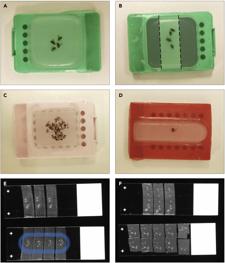
Formalin-Fixed Paraffin-Embedded blocks and sections (A and B) Correct positioning of fruit flies in the paraffin block. The gray area (B) shows the excess of paraffin to trim with a scalpel before sectioning in order to place more sections on one slide. (C) A paraffin block with too many fruit flies, some appears damaged. (D) Fruit fly embedded in a rectangular mold (less paraffin to trim). (E) Correct placement of sections on a slide to be processed for immunofluorescence staining. The blue circle on the bottom slide shows how the hydrophilic border can be drawn after deparaffinization. During immunostaining, solution (steps 49–51, 53, 55 and 56) should be added within the circle. (F) Placement suitable for visualization without processing but fruit flies are too close to the edge of the slide to draw a hydrophilic border for a staining procedure.

Bright field observation of unprocessed section of a fruit fly (A) 4 μm section of a fruit fly without biofilm. Fruit fly body is divided into three anatomical parts: head, thorax, and abdomen. (B) Fruit fly infected with P. acnes biofilm. (C) Close-up of figure A. Scale bar: 500 μm.

Immunofluorescence of in vivo Propionibacterium acnes biofilm (A and B) P. acnes is labeled with anti-P. acnes monoclonal antibody/Alexa Fluor® 555 goat anti-mouse IgG. Host and bacterial nuclei are stained with DAPI. Arrows highlight the fruit fly gut wall. Scale bar: 20 μm.

Immunofluorescence of a non-germ-free Drosophila melanogaster infected for three days with Propionibacterium acnes (A and B) Sections of (A) WT and (B) contaminated GF fruit flies are stained with DAPI and anti-P. acnes monoclonal antibody/Alexa Fluor® 488 goat anti-mouse IgG. The presence of rod-shaped bacteria other than P. acnes is visible in the merge image. Scale bar: 20 μm.
References
-
- BDSC Bloomington Drosophila Stock Center, Fly Foods & Methods - Semi-defined Food. 2019. https://bdsc.indiana.edu/information/recipes/germanfood.html
-
- Bronnec V., Eilers H., Jahns A.C., Omer H., Alexeyev O.A. Propionibacterium (Cutibacterium) granulosum extracellular DNase BmdE targeting Propionibacterium (Cutibacterium) acnes biofilm matrix, a novel inter-species competition mechanism. Front. Cell. Infect. Microbiol. 2022;1337 doi: 10.3389/fcimb.2021.809792. - DOI - PMC - PubMed
-
- Chauhan A., Jindal T. Microbiological Methods for Environment, Food and Pharmaceutical Analysis. Springer; 2020. Good microbiological laboratory practices; pp. 15–22. - DOI
-
- CSHProtocols Cold spring harbor protocols: recipe 10X PBS. 2007. http://cshprotocols.cshlp.org/content/2007/4/pdb.rec10768.full?text_only...
Publication types
MeSH terms
LinkOut - more resources
Full Text Sources
Molecular Biology Databases
Miscellaneous
