. 2022 Apr 15:16:797588.
doi: 10.3389/fncel.2022.797588. eCollection 2022.

Plasticity-Related Gene 5 Is Expressed in a Late Phase of Neurodifferentiation After Neuronal Cell-Fate Determination

Affiliations

Plasticity-Related Gene 5 Is Expressed in a Late Phase of Neurodifferentiation After Neuronal Cell-Fate Determination

Isabel Gross et al. Front Cell Neurosci. .

Abstract

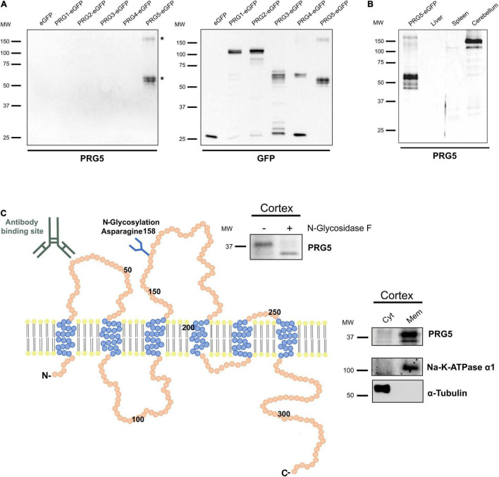
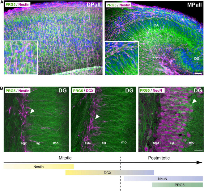
During adult neurogenesis, neuronal stem cells differentiate into mature neurons that are functionally integrated into the existing network. One hallmark during the late phase of this neurodifferentiation process is the formation of dendritic spines. These morphological specialized structures form the basis of most excitatory synapses in the brain, and are essential for neuronal communication. Additionally, dendritic spines are affected in neurological disorders, such as Alzheimer's disease or schizophrenia. However, the mechanisms underlying spinogenesis, as well as spine pathologies, are poorly understood. Plasticity-related Gene 5 (PRG5), a neuronal transmembrane protein, has previously been linked to spinogenesis in vitro. Here, we analyze endogenous expression of the PRG5 protein in different mouse brain areas, as well as on a subcellular level. We found that native PRG5 is expressed dendritically, and in high abundance in areas characterized by their regenerative capacity, such as the hippocampus and the olfactory bulb. During adult neurogenesis, PRG5 is specifically expressed in a late phase after neuronal cell-fate determination associated with dendritic spine formation. On a subcellular level, we found PRG5 not to be localized at the postsynaptic density, but at the base of the synapse. In addition, we showed that PRG5-induced formation of membrane protrusions is independent from neuronal activity, supporting a possible role in the morphology and stabilization of spines.

Keywords: PRGs; brain development; dendritic spines; neurodifferentiation; neurogenesis.


Conflict of interest statement

The authors declare that the research was conducted in the absence of any commercial or financial relationships that could be construed as a potential conflict of interest.

Figures

FIGURE 1
FIGURE 1
PRG5 is a glycosylated membrane protein in brain tissue. (A) Specificity analysis of a polyclonal PRG5 antibody shows specific binding only to PRG5 and no cross-reactivity to other PRG family members. Fusion proteins of PRG1-5 with eGFP were used for western blotting. The PRG5 antibody detected bands around 50 kDa and above 150 kDa. Bands marked with asterisks (*) were analyzed by MS and contained PRG5 peptides. (B) Western Blot analysis of protein lysates of adult mouse tissue shows strong PRG5 signal in the cerebellum, but not in liver or spleen (probed with anti-PRG5 antibody). Loading control is shown in Supplementary Figure 1. (C) Schematic depiction of the murine PRG5 protein and its orientation in the plasma membrane based on its amino acid sequence (primary accession number Q8BJ52). The six transmembrane regions are illustrated in blue, N- and C-termini are located intracellularly. The antibody-binding site in the first extracellular loop is indicated. An N-glycosylation site at asparagine 158 in the second extracellular loop is indicated, and was supported by western blot of P15 mouse cortex lysate treated with (+) and without (–) N-glycosidase F, and a subsequent shift of the PRG5 band to a lower molecular weight after treatment (blot above). Endogenous membrane localization of PRG5 was validated by western blot of cytosolic (Cyt) and membrane (Mem) protein fractions of P15 mouse cortex lysates (blot on the right). α-tubulin was used as a cytosolic marker, Na-K-ATPase α1 as a membrane marker. MW, molecular weight.
FIGURE 2
FIGURE 2
PRG5 protein expression in the mouse cortex increases during maturation. (A) Representative western blot analysis of total protein lysates of mouse cortex tissue between E14 and P60, probed with an anti-PRG5 antibody. The 150 kDa band was detected from E19 and increased toward adult stages. The 37 kDa band was detectable from P10. Only a very weak band was detectable in younger stages. Ponceau-S staining of total proteins is shown as a loading control. A representative blot of three technical repeats for each of three tissue preparations is shown. MW, molecular weight. (B) Immunostaining of PRG5 (green) and DAPI (blue) in a sagittal section of P15 mouse cortex. PRG5 expression was detected in neurons of all cortical layers. Asterisk (*) indicates pial surface. Scale bar = 100 μm.
FIGURE 3
FIGURE 3
PRG5 is enriched in dendritic areas of the mouse hippocampus, olfactory bulb, and cerebellum. (A) Montage of a sagittal adult mouse brain section labeled with PRG5 (green) shows highest PRG5 expression in the cerebellum, hippocampus, and the olfactory bulb. Mosaic stitched from 635 tiles. Scale bar = 1 mm. (B) Dendritic expression of PRG5 (green) in the hippocampal CA1 region shown by co-immunostaining with MAP2 (magenta). PRG5 expression was found in the soma of pyramidal cells of the sp and increased in their dendritic arbors in the sr and slm. Scale bar = 100 μm. (C) Low axonal expression of PRG5 in the DG of the hippocampus is shown by co-immunostaining with axonal marker NF-M (magenta). Strong PRG5 expression was found in granule cells in the sg and their dendritic arbors in the mo, whereas there was only low expression in their mossy fibers in the hilum. Scale bar = 10 μm. (D) Purkinje cells of the CB showed strong PRG5 signal in their soma and in their dendritic arbors in the mo. Unspecific overlapping staining of blood vessels is marked with asterisks. Scale bar = 20 μm. (E) NeuN positive granule cells of the granule layer of the CB show no PRG5 expression. Scale bar = 20 μm. DAPI visualizes cell nuclei (blue). CA, cornu ammonis; CB, cerebellum; CTX, cerebral cortex; DG, dentate gyrus; gl, glomerular layer; gr, granule layer; h, hilus; HB, hindbrain; HPF, hippocampus formation; HY, hypothalamus; ipl, inner plexiform layer; MB, midbrain; mi, mitral layer; mo, molecular layer; MOB, main olfactory bulb; opl, outer plexiform layer; pu, Purkinje layer; sg, stratum granulosum; sgz, subgranular zone; slm, stratum lacunosum-moleculare; so, stratum oriens; sp, stratum pyramidale; sr, stratum radiale; STR, striatum; TH, thalamus. Representative images; Stainings of brain sections from at least three different animals were analyzed and revealed similar results.
FIGURE 4
FIGURE 4
Cell-type specific expression of PRG5 in the brain. (A) TH-positive (magenta) dopaminergic neurons in the gl of the MOB expressed PRG5 (green). Scale bar = 20 μm. (B) PV (magenta)-positive inhibitory GABAergic interneurons in the hippocampus also expressed PRG5 (green). Scale bar = 20 μm. (C) PRG5 (green) was not expressed by GFAP-positive (magenta) astrocytes. Scale bar = 20 μm. (D) NF-M-expressing (magenta) axonal fibers in the cc did not show PRG5 (green) positive staining, but a PRG5-expressing subtype of oligodendrocytes was detected. Scale bar = 20 μm. DAPI was used to visualize cell nuclei (blue). CA, cornu ammonis; cc, corpus callosum; MOB, main olfactory bulb. Representative images; Stainings of brain sections from at least three different animals were analyzed and revealed similar results.
FIGURE 5
FIGURE 5
PRG5 protein is expressed after neuronal cell fate determination. (A) Representative sagittal sections of P0 mice brain, stained with antibodies against PRG5 (green) and nestin (magenta), revealed PRG5 expression in the developing isocortex (dorsal pallium, DPall) and the developing hippocampus (medial pallium, MPall) that was not colocalized to nestin-positive progenitor cells. Scale bar = 50 μm. (B) During adult neurogenesis in the sgz of the dentate gyrus in the hippocampus, PRG5 (green) expression was detected in a late postmitotic phase, together with NeuN (magenta, right image) expression, but not with mitotic neurodifferentiation markers nestin (magenta, left image) and DCX (magenta, middle image). Scale bar = 20 μm. CA, cornu ammonis; DG, dentate gyrus; DPall, dorsal pallium; mo, molecular layer, MPall, medial pallium; sg, stratum granulosum; sgz, subgranular zone. Representative images; Stainings of brain sections from at least three different animals were analyzed and revealed similar results.
FIGURE 6
FIGURE 6
PRG5 protein expression is present in a late phase of neurodifferentiation in adult neurogenesis of the rostral migratory stream (RMS). Top: Montage of a sagittal adult mouse brain section labeled with PRG5 (green), DCX (magenta) and DAPI (cell nuclei, blue). Mosaic stitched from 234 tiles. Bottom: Full-resolution views of representative stages of migration in the RMS and schematic drawing of the svz, RMS, and the olfactory bulb in sagittal view. PRG5 protein abundance was only present at a late stage of neurodifferentiation, when neuroblasts entered the olfactory bulb and started differentiating. HPF, hippocampal formation; MOB, main olfactory bulb; RMS, rostral migratory stream, svz, subventricular zone; VL, lateral ventricle. Scale bar = 50 μm. Representative images; Stainings of brain sections from at least three different animals were analyzed and revealed similar results.
FIGURE 7
FIGURE 7
PRG5 stimulates formation of dendritic protrusions, independent of neuronal activity. (A) Representative western blot after subcellular fractionation of adult mouse cerebral cortex, probed with PRG5, with PSD95 as a postsynaptic density marker, HSP60 as a mitochondrial marker, synaptophysin as a synaptic marker, and MBP as a myelin marker. PRG5 was detected in myelin-, ER-, Golgi-enriched, and in synaptosomal fractions, predominantly around 150 kDa. The 37 kDa PRG5 band was mainly detected in the myelin-enriched and the crude synaptic plasma-membrane fractions. No PRG5 signal was found in the postsynaptic density fraction. A representative blot of three technical repeats for each of three tissue preparations is shown. MW, molecular weight. (B) Representative volume-rendered 3D projections of z-stack images of murine primary neurons cultured 15 days in vitro and stained for PRG5 (green), homer1 (magenta), and bassoon (top, blue) or MAP2 (bottom, blue). Stainings were analyzed for three independent neuron preparations. PRG5 protein was not localized to pre- and postsynaptic transmission sites, as shown by a lack of signal overlap to homer1 or bassoon proteins (top, white arrow heads). PRG5 was localized at the bases of postsynaptic spines, between postsynaptic density protein homer1 and dendritic-cytoskeletal protein MAP2 (bottom, white arrow heads). Scale bars = 1 μm. (C) Quantification of protrusions of mature primary neurons transfected either with CFP-MEM (control) or PRG5-eGFP with (+) and without (–) activity inhibition (1 μM TTX, 50 μM D-APV and 10 μM nifedipine). PRG5- overexpressing neurons showed elevated number of overall protrusions and of homer-positive protrusions compared to control cells after inhibition of neuronal activity. Number of protrusions was normalized to control (CFP-MEM) without activity inhibition (100%) and are shown as mean in% + SEM. Counted numbers and n-values are listed in the table below graphs. Statistical data is shown in Supplementary Table 2.

Similar articles

Cited by

References

    1. Altman J., Das G. D. (1965). Autoradiographic and histological evidence of postnatal hippocampal neurogenesis in rats. J. Comp. Neurol. 124 319–335. 10.1002/cne.901240303 - DOI - PubMed
    1. Brandt N., Franke K., Rasin M. R., Baumgart J., Vogt J., Khrulev S., et al. (2007). The neural EGF family member CALEB/NGC mediates dendritic tree and spine complexity. Embo J. 26 2371–2386. 10.1038/sj.emboj.7601680 - DOI - PMC - PubMed
    1. Bräuer A. U., Nitsch R. (2008). Plasticity-related genes (PRGs/LRPs): a brain-specific class of lysophospholipid-modifying proteins. Biochim. Biophys. Acta 1781 595–600. 10.1016/j.bbalip.2008.04.004 - DOI - PubMed
    1. Bräuer A. U., Savaskan N. E., Kuhn H., Prehn S., Ninnemann O., Nitsch R. (2003). A new phospholipid phosphatase, PRG-1, is involved in axon growth and regenerative sprouting. Nat. Neurosci. 6 572–578. 10.1038/nn1052 - DOI - PubMed
    1. Brewer G. J., Torricelli J. R., Evege E. K., Price P. J. (1993). Optimized survival of hippocampal neurons in B27-supplemented Neurobasal, a new serum-free medium combination. J. Neurosci. Res. 35 567–576. 10.1002/jnr.490350513 - DOI - PubMed

LinkOut - more resources