

Primary Myelofibrosis in the Prefibrotic Stage Presenting as Portal, Splenic, and Superior Mesenteric Vein Thrombosis: A Case Report and Review of the Literature
- PMID: 35497670
- PMCID: PMC8995663
- DOI: 10.1159/000514658
Primary Myelofibrosis in the Prefibrotic Stage Presenting as Portal, Splenic, and Superior Mesenteric Vein Thrombosis: A Case Report and Review of the Literature
Abstract
Introduction: Myeloproliferative neoplasms are the most common cause of splanchnic vein thrombosis in the absence of cirrhosis or nearby malignancy.
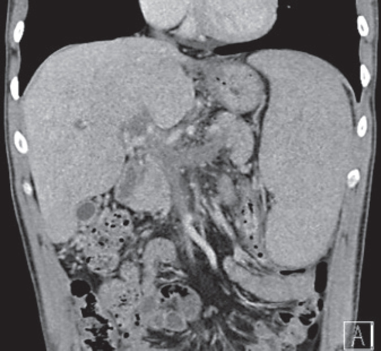
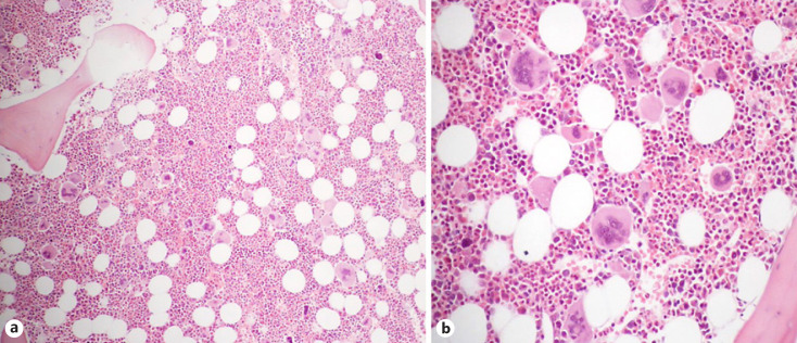
Case presentation: A 31-year-old male presented to the emergency department with epigastric pain associated with mild thrombocytosis and elevated levels of aminotransferases, lactate dehydrogenase, and C-reactive protein. Contrast-enhanced abdominal computed tomography revealed splanchnic venous thrombosis that involved the portal, splenic, and superior mesenteric veins, without signs of chronic liver disease. Anticoagulation with warfarin was immediately started. Diagnostic work-up was remarkable for the presence of the JAK2 V617T mutation and hypercellular bone marrow, with increased myeloid cells and atypical megakaryocytes, consistent with primary myelofibrosis in a prefibrotic stage. No other hypercoagulable conditions were identified.
Discussion: We present a rare case of primary myelofibrosis in the prefibrotic stage presenting as portal-splenic-superior mesenteric vein thrombosis. This demonstrates that extensive splanchnic vein thrombosis may be the onset manifestation of myeloproliferative neoplasms, even in early stages and in the absence of concomitant hypercoagulable conditions. The presence of the JAK2 mutation is an important prothrombotic risk factor that can, per se, contribute to large venous thrombosis.
Introdução: As neoplasias mieloproliferativas constituem a causa mais comum de trombose venosa esplâncnica na ausência de cirrose hepatica ou neoplasia regional.
Descrição do caso: Um homem de 31 anos apresentou-se no Serviço de Urgência com dor epigástrica associada a trombocitose ligeira e elevação das transamínases, desidrogenase láctica e proteína C-reactiva. Em tomografia computorizada abdominal com contraste, foi identificada trombose venosa esplâncnica envolvendo a veia porta, esplénica e mesentérica superior, sem sinais de doença hepática crónica. Foi de imediato iniciada anticoagulação com varfarina. Da investigação etiológica, destaca-se a presença da mutação JAK2 V617F e medula óssea hiper-celular com aumento das contagens de células mielóides e megacariócitos atípicos, consistente com mielofibrose primária em estadio pré-fibrótico. Não se identificaram distúrbios pro-trombóticos concomitantes.
Discussão: Apresenta-se um raro caso de trombose da veia porta, esplénica e mesentérica superior, demonstrando que as neoplasias mieloproliferativas podem apresentar-se sob a forma de trombose venosa esplâncnica extensa, mesmo em estadios precoces e na ausência de distúrbios protrombóticos concomitantes. A presença da mutação JAK2 é um importante factor de risco pro-trombótico que pode por si só contribuir para a formação de tromboses venosas extensas.
Keywords: Myeloproliferative neoplasm; Portal vein thrombosis; Primary myelofibrosis; Splanchnic vein thrombosis.
Copyright © 2021 by S. Karger AG, Basel.
Conflict of interest statement
The authors have no conflicts of interest to declare.
Figures
References
-
- Rajani R, Björnsson E, Bergquist A, Danielsson A, Gustavsson A, Grip O, et al. The epidemiology and clinical features of portal vein thrombosis: a multicentre study. Aliment Pharmacol Ther. 2010 Nov;32((9)):1154–62. - PubMed
-
- Smalberg JH, Arends LR, Valla DC, Kiladjian JJ, Janssen HL, Leebeek FW. Myeloproliferative neoplasms in Budd-Chiari syndrome and portal vein thrombosis: a meta-analysis. Blood. 2012 Dec;120((25)):4921–8. - PubMed
-
- Lavu S, Szuber N, Mudireddy M, Yogarajah M, Gangat N, Pardanani A, et al. Splanchnic vein thrombosis in patients with myeloproliferative neoplasms: the Mayo clinic experience with 84 consecutive cases. Am J Hematol. 2018 Mar;93((3)):E61–4. - PubMed
Publication types
LinkOut - more resources
Full Text Sources
Research Materials
Miscellaneous
