. 2021 Dec 16;7(4):797-809.
doi: 10.1016/j.ekir.2021.11.037. eCollection 2022 Apr.

Minimal Change Disease Is Associated With Endothelial Glycocalyx Degradation and Endothelial Activation

Affiliations

Minimal Change Disease Is Associated With Endothelial Glycocalyx Degradation and Endothelial Activation

Colin Bauer et al. Kidney Int Rep. .

Abstract

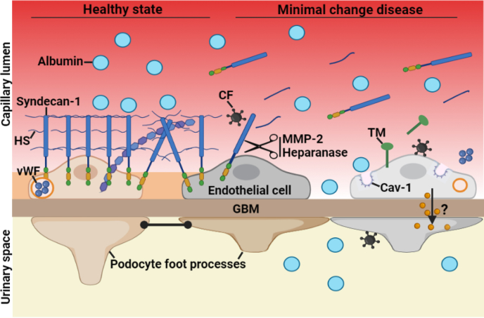
Introduction: Minimal change disease (MCD) is considered a podocyte disorder triggered by unknown circulating factors. Here, we hypothesized that the endothelial cell (EC) is also involved in MCD.

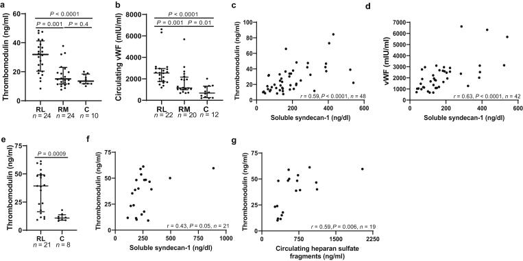
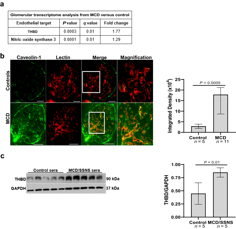
Methods: We studied 45 children with idiopathic nephrotic syndrome (44 had steroid sensitive nephrotic syndrome [SSNS], and 12 had biopsy-proven MCD), 21 adults with MCD, and 38 healthy controls (30 children, 8 adults). In circulation, we measured products of endothelial glycocalyx (EG) degradation (syndecan-1, heparan sulfate [HS] fragments), HS proteoglycan cleaving enzymes (matrix metalloprotease-2 [MMP-2], heparanase activity), and markers of endothelial activation (von Willebrand factor [vWF], thrombomodulin) by enzyme-linked immunosorbent assay (ELISA) and mass spectrometry. In human kidney tissue, we assessed glomerular EC (GEnC) activation by immunofluorescence of caveolin-1 (n = 11 MCD, n = 5 controls). In vitro, we cultured immortalized human GEnC with sera from control subjects and patients with MCD/SSNS sera in relapse (n = 5 per group) and performed Western blotting of thrombomodulin of cell lysates as surrogate marker of endothelial activation.

Results: In circulation, median concentrations of all endothelial markers were higher in patients with active disease compared with controls and remained high in some patients during remission. In the MCD glomerulus, caveolin-1 expression was higher, in an endothelial-specific pattern, compared with controls. In cultured human GEnC, sera from children with MCD/SSNS in relapse increased thrombomodulin expression compared with control sera.

Conclusion: Our data show that alterations involving the systemic and glomerular endothelium are nearly universal in patients with MCD and SSNS, and that GEnC can be directly activated by circulating factors present in the MCD/SSNS sera during relapse.

Keywords: endothelial activation; endothelial glycocalyx; glomerular endothelial cell; minimal change disease; podocyte; steroid sensitive nephrotic syndrome.


Figures

None
Graphical abstract
Figure 1
Figure 1
Endothelial glycocalyx degradation in children with MCD and SSNS. Datapoints indicate the average of duplicate measurements per subject of the following markers: (a) circulating syndecan-1; (b) circulating HS fragments; (c) correlation between syndecan-1 and HS fragments; (d) circulating MMP-2; (e) correlation between MMP-2 and syndecan-1; (f) heparanase activity in circulation; (g) correlation between heparanase activity and syndecan-1. Data presented as median ± interquartile range. Outside values were included in analyses and were a total of 3 for syndecan-1 (1 RL, 1 RM, 1 C), 1 for HS (relapse), 1 for MMP-2 (RM), and 2 for heparanase (RL). C, control; HS, heparan sulfate; MCD, minimal change disease; MMP-2, matrix metalloprotease-2; RL, relapse; RM, remission; SSNS, steroid sensitive nephrotic syndrome.
Figure 2
Figure 2
Endothelial glycocalyx degradation in adults with MCD. Datapoints indicate the average of duplicate measurements per subject of the following markers: (a) circulating syndecan-1; (b) circulating HS; (c) correlation between circulating syndecan-1 and HS. Data presented as median ± interquartile range. Outside values were included in analyses and were a total of 1 for syndecan-1 and heparan sulfate (RL, for both groups). C, control; HS, heparan sulfate; MCD, minimal change disease; RL, relapse.
Figure 3
Figure 3
MCD is associated with systemic endothelial activation. Datapoints indicate the average of duplicate measurements per subject of the following markers: (a) circulating thrombomodulin in children; (b) circulating vWF in children; (c) correlation between thrombomodulin and syndecan-1 in children; (d) correlation between vWF and syndecan-1 in children; (e) circulating thrombomodulin in adults; (f) correlation between syndecan-1 and thrombomodulin; (g) correlation between HS and thrombomodulin in adults. Data presented as median ± interquartile range. Outside values were included in analyses and were a total of 2 for thrombomodulin (2 RM) and 3 for vWF (2 RL, 1 RM) from the pediatric cohort. C, control; HS, heparan sulfate; MCD, minimal change disease; RL, relapse; RM, remission; vWF, von Willebrand factor.
Figure 4
Figure 4
Glomerular endothelial activation in MCD and SSNS. (a) Glomerular transcriptome analysis from patients with MCD in relapse versus controls obtained from Nephroseq.org (“Ju CKD Glom cohort”); (b) immunofluorescence of human kidney showed up-regulation of caveolin-1 (green), colocalizing with rhodamine lectin (red), in adults with MCD in relapse compared with controls (n = 11 vs. n = 5), scale bars = 100 μm. Right graph represents immunofluorescence analysis of caveolin-1 expression in human kidney tissue, P = 0.0005; (c) Western blots of THBD and GAPDH from hiGEnC exposed for 24 hours to 10% sera from children with MCD/SSNS in relapse (n = 1/n = 4, respectively) versus control subjects (n = 5). Right graph represents densitometry analysis of Western blots from hiGEnC lysates, P = 0.01. Data presented as median ± interquartile range. GAPDH, Glyceraldehyde 3-phosphate dehydrogenase; hiGEnC, human immortalized glomerular endothelial cell; MCD, minimal change disease; SSNS, steroid sensitive nephrotic syndrome; THBD, thrombomodulin.
Figure 5
Figure 5
Schematic showing endothelial alterations in MCD. In healthy state, the EG, which predominantly consists of syndecan-1 and HS, covers the luminal surface of the glomerular endothelial cells and their fenestrations. In MCD, the CF directly targets the endothelium, resulting in EG degradation, as noted by the shedding of syndecan-1 and HS into circulation, and GEnC activation involving the release of TM and vWF into circulation, and the up-regulation of Cav-1. We postulate that EG degradation may favor the passage of serum albumin through endothelial fenestration and may facilitate that circulating factors encounter and activate podocytes. Furthermore, activated GEnC may release factors locally that could contribute to podocyte injury. Cartoon created with BioRender.com. Cav-1, caveolin-1; CF, circulating factor; EG, endothelial glycocalyx; GBM, glomerular basement membrane; GEnC, glomerular endothelial cell; HS, heparan sulfate; MCD, minimal change disease; MMP-2, matrix metalloprotease-2; TM, thrombomodulin; vWF, von Willebrand factor.

References

    1. Vivarelli M., Massella L., Ruggiero B., Emma F. Minimal change disease. Clin J Am Soc Nephrol. 2017;12:332–345. doi: 10.2215/CJN.05000516. - DOI - PMC - PubMed
    1. Kopp J.B., Anders H.J., Susztak K., et al. Podocytopathies. Nat Rev Dis Primers. 2020;6:68. doi: 10.1038/s41572-020-0196-7. - DOI - PMC - PubMed
    1. Colucci M., Corpetti G., Emma F., Vivarelli M. Immunology of idiopathic nephrotic syndrome. Pediatr Nephrol. 2018;33:573–584. doi: 10.1007/s00467-017-3677-5. - DOI - PubMed
    1. Shalhoub R.J. Pathogenesis of lipoid nephrosis: a disorder of T-cell function. Lancet. 1974;2:556–560. doi: 10.1016/s0140-6736(74)91880-7. - DOI - PubMed
    1. Grahammer F., Schell C., Huber T.B. The podocyte slit diaphragm—from a thin grey line to a complex signalling hub. Nat Rev Nephrol. 2013;9:587–598. doi: 10.1038/nrneph.2013.169. - DOI - PubMed

LinkOut - more resources