

Clinicopathologic and Prognostic Study of Primary IgA Nephropathy With Light Chain λ Restriction in the Mesangial Deposits
- PMID: 35497802
- PMCID: PMC9039423
- DOI: 10.1016/j.ekir.2022.01.1053
Clinicopathologic and Prognostic Study of Primary IgA Nephropathy With Light Chain λ Restriction in the Mesangial Deposits
Abstract
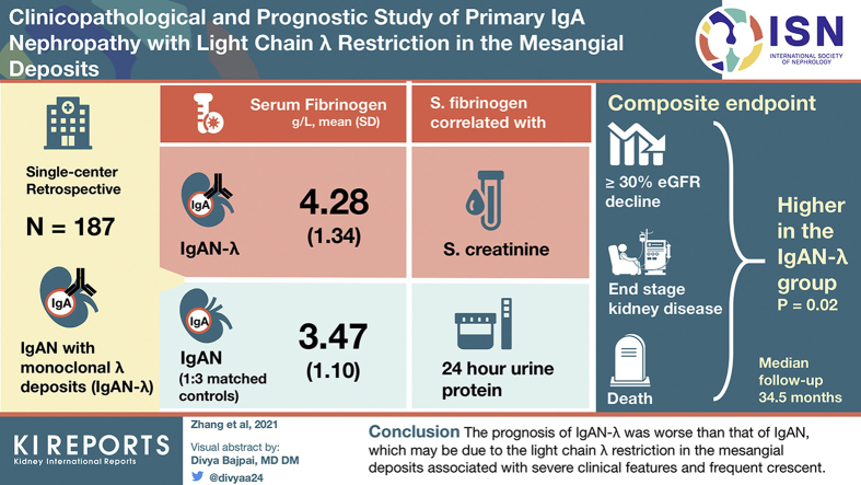
Introduction: Primary IgA nephropathy (IgAN) with light chain λ restriction in the mesangial deposits (IgAN-λ) has unique immunofluorescence (IF) features. Nevertheless, its clinicopathology and prognosis are still ambiguous.
Methods: From January 2002 to December 2020, the clinical and pathologic data of 3872 patients who were diagnosed with having primary IgAN by renal biopsy in our hospital were reviewed. A total of 187 patients who met the selection criteria for IgAN-λ were enrolled to conduct a retrospective single-center study. The selection criteria were that IF features conform to light chain λ restriction in the mesangial deposits. According to age, sex, renal function (estimated glomerular filtration rate [eGFR]), and follow-up time, the control group was constructed with 1:3 matched cases of IgAN. The clinicopathologic and prognostic differences between the 2 groups were analyzed.
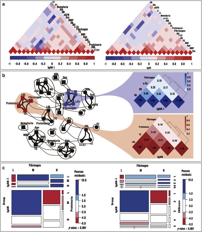
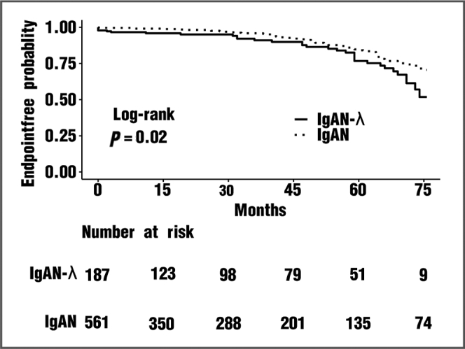
Results: Compared with that in the IgAN group, the serum fibrinogen level in the IgAN-λ group was significantly higher (P < 0.001). Furthermore, cluster analysis indicated the different clusters involved in fibrinogen between the IgAN-λ and IgAN groups and that fibrinogen is associated with factors reflecting renal function in IgAN-λ but proteinuria levels in IgAN. The light chain λ deposit in the mesangium is associated with the formation of crescents in those with IgAN-λ, but complement C3 deposition in those with IgAN. Our Kaplan-Meier analysis revealed that the prognosis of the IgAN-λ group was significantly worse than that of the IgAN group within >6 years of follow-up (P = 0.02). The multi-Cox analysis revealed that the light chain λ restriction in the mesangial deposits was an independent risk factor for poor outcomes (eGFR decreased from the baseline ≥ 30% continuously or reached end-stage renal disease [ESRD] or died).
Conclusion: The prognosis of those with IgAN-λ was worse than that of those with IgAN, which may be attributed to the light chain λ restriction in the mesangial deposits inducing a significant systemic inflammation manifested as severe clinical features and frequent crescent.
Keywords: IgA nephropathy; clinicopathology; kidney disease; light chain λ; prognosis.
© 2022 International Society of Nephrology. Published by Elsevier Inc.
Figures
Similar articles
-
Immunofluorescence deposits in the mesangial area and glomerular capillary loops did not affect the prognosis of immunoglobulin a nephropathy except C1q:a single-center retrospective study.BMC Nephrol. 2021 Jan 29;22(1):43. doi: 10.1186/s12882-021-02237-w. BMC Nephrol. 2021. PMID: 33514328 Free PMC article.
-
Clinical and prognostic significance of C1q deposition in IgAN patients-a retrospective study.Int Immunopharmacol. 2020 Nov;88:106896. doi: 10.1016/j.intimp.2020.106896. Epub 2020 Oct 14. Int Immunopharmacol. 2020. PMID: 33182045
-
Roles of mesangial C3 and C1q deposition in the clinical manifestations and prognosis of IgAN.Int Immunopharmacol. 2023 Jul;120:110354. doi: 10.1016/j.intimp.2023.110354. Epub 2023 May 24. Int Immunopharmacol. 2023. PMID: 37235963
-
Proliferative glomerulonephritis with monoclonal immunoglobulin G deposits complicated by immunoglobulin A nephropathy in the renal allograft.Nephrology (Carlton). 2016 Jul;21 Suppl 1:48-52. doi: 10.1111/nep.12775. Nephrology (Carlton). 2016. PMID: 26971743 Review.
-
Factors predicting progression of IgA nephropathies.J Nephrol. 2005 Sep-Oct;18(5):503-12. J Nephrol. 2005. PMID: 16299675 Review.
Cited by
-
Clinicopathological and prognostic study of idiopathic membranous nephropathy with tip or non-tip variant focal segmental glomerulosclerosis: a single-center cohort study.Ren Fail. 2025 Dec;47(1):2512052. doi: 10.1080/0886022X.2025.2512052. Epub 2025 Jun 8. Ren Fail. 2025. PMID: 40484026 Free PMC article.
-
Colocalization of IgG and IgA Heavy Chains with Kappa and Lambda Light Chains in Glomerular Deposits of IgA Nephropathy Patients Using High-Resolution Confocal Microscopy and Correlation with Oxford MEST-C Scores.J Clin Med. 2023 Nov 28;12(23):7361. doi: 10.3390/jcm12237361. J Clin Med. 2023. PMID: 38068413 Free PMC article.
-
Clinicopathologic Significance of Predominant Lambda Light Chain Deposition in IgA Nephropathy.Kidney Int Rep. 2022 Aug 17;7(11):2462-2473. doi: 10.1016/j.ekir.2022.08.003. eCollection 2022 Nov. Kidney Int Rep. 2022. PMID: 36531879 Free PMC article.
References
LinkOut - more resources
Full Text Sources
Research Materials
Miscellaneous
