

The Endothelium as a Hub for Cellular Communication in Atherogenesis: Is There Directionality to the Message?
- PMID: 35498030
- PMCID: PMC9051343
- DOI: 10.3389/fcvm.2022.888390
The Endothelium as a Hub for Cellular Communication in Atherogenesis: Is There Directionality to the Message?
Abstract
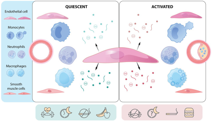
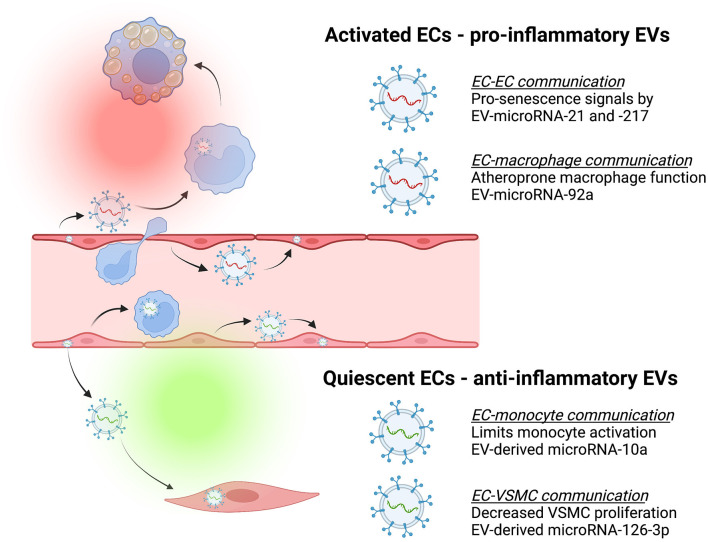
Endothelial cells line every blood vessel and thereby serve as an interface between the blood and the vessel wall. They have critical functions for maintaining homeostasis and orchestrating vascular pathogenesis. Atherosclerosis is a chronic disease where cholesterol and inflammatory cells accumulate in the artery wall below the endothelial layer and ultimately form plaques that can either progress to occlude the lumen or rupture with thromboembolic consequences - common outcomes being myocardial infarction and stroke. Cellular communication lies at the core of this process. In this review, we discuss traditional (e.g., cytokines, chemokines, nitric oxide) and novel (e.g., extracellular vesicles) modes of endothelial communication with other endothelial cells as well as circulating and vessel wall cells, including monocytes, macrophages, neutrophils, vascular smooth muscle cells and other immune cells, in the context of atherosclerosis. More recently, the growing appreciation of endothelial cell plasticity during atherogenesis suggests that communication strategies are not static. Here, emerging data on transcriptomics in cells during the development of atherosclerosis are considered in the context of how this might inform altered cell-cell communication. Given the unique position of the endothelium as a boundary layer that is activated in regions overlying vascular inflammation and atherosclerotic plaque, there is a potential to exploit the unique features of this group of cells to deliver therapeutics that target the cellular crosstalk at the core of atherosclerotic disease. Data are discussed supporting this concept, as well as inherent pitfalls. Finally, we briefly review the literature for other regions of the body (e.g., gut epithelium) where cells similarly exist as a boundary layer but provide discrete messages to each compartment to govern homeostasis and disease. In this light, the potential for endothelial cells to communicate in a directional manner is explored, along with the implications of this concept - from fundamental experimental design to biomarker potential and therapeutic targets.
Keywords: atherosclerosis; crosstalk; directionality; endothelium; extracellular vesicles; inflammation; microRNA; polarity.
Copyright © 2022 Howe, Cybulsky and Fish.
Conflict of interest statement
The authors declare that the research was conducted in the absence of any commercial or financial relationships that could be construed as a potential conflict of interest.
Figures
References
Publication types
LinkOut - more resources
Full Text Sources