RAGE Regulating Vascular Remodeling in Diabetes by Regulating Mitochondrial Dynamics with JAK2/STAT3 Pathway
- PMID: 35498181
- PMCID: PMC9054424
- DOI: 10.1155/2022/2685648
RAGE Regulating Vascular Remodeling in Diabetes by Regulating Mitochondrial Dynamics with JAK2/STAT3 Pathway
Retraction in
-
Retracted: RAGE Regulating Vascular Remodeling in Diabetes by Regulating Mitochondrial Dynamics with JAK2/STAT3 Pathway.Comput Intell Neurosci. 2023 Jul 26;2023:9832645. doi: 10.1155/2023/9832645. eCollection 2023. Comput Intell Neurosci. 2023. PMID: 37538730 Free PMC article.
Abstract
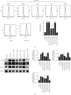
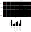
In this research, we will explore the role and modulation of mitochondrial dynamics in diabetes vascular remodeling. Only a few cell types express the pattern recognition receptor, also known as the AGE receptor (RAGE). However, it is triggered in almost all of the cells that have been investigated thus far by events that are known to cause inflammation. Here, Type 2 diabetes was studied in both cellular and animal models. Elevated Receptor for advanced glycation end products (RAGE), phosphorylated JAK2 (p-JAK2), phosphorylated STAT3 (p-STAT3), transient receptor potential ion channels (TRPM), and phosphorylated dynamin-related protein 1 (p-DRP1) were observed in the context of diabetes. In addition, we found that inhibition of RAGE was followed by a remarkable decrease in the expression of the above proteins. It has also been demonstrated by western blotting and immunofluorescence results in vivo and in vitro. Suppressing STAT3 and DRP1 phosphorylation produced effects similar to those of RAGE inhibition on the proliferation, cell cycle, migration, invasion, and expression of TRPM in VSMCs and vascular tissues obtained from diabetic animals. These findings indicate that RAGE regulates vascular remodeling via mitochondrial dynamics through modulating the JAK2/STAT3 axis in diabetes. The findings could be crucial in gaining a better understanding of diabetes-related vascular remodeling. It also contributes to a better cytopathological understanding of diabetic vascular disease and provides a theoretical foundation for novel targets that aid in the prevention and treatment of diabetes-related cardiovascular problems.
Copyright © 2022 Shengjia Sun et al.
Conflict of interest statement
The authors confirm that there are no conflicts of interest.
Figures


Similar articles
-
RAGE-dependent activation of the oncoprotein Pim1 plays a critical role in systemic vascular remodeling processes.Arterioscler Thromb Vasc Biol. 2011 Sep;31(9):2114-24. doi: 10.1161/ATVBAHA.111.230573. Epub 2011 Jun 16. Arterioscler Thromb Vasc Biol. 2011. PMID: 21680901 Free PMC article.
-
Profilin-1 is involved in macroangiopathy induced by advanced glycation end products via vascular remodeling and inflammation.World J Diabetes. 2021 Nov 15;12(11):1875-1893. doi: 10.4239/wjd.v12.i11.1875. World J Diabetes. 2021. PMID: 34888013 Free PMC article.
-
Advanced glycation end products receptor RAGE controls myocardial dysfunction and oxidative stress in high-fat fed mice by sustaining mitochondrial dynamics and autophagy-lysosome pathway.Free Radic Biol Med. 2017 Nov;112:397-410. doi: 10.1016/j.freeradbiomed.2017.08.012. Epub 2017 Aug 19. Free Radic Biol Med. 2017. PMID: 28826719
-
Advanced Glycation End Products and Diabetes Mellitus: Mechanisms and Perspectives.Biomolecules. 2022 Apr 4;12(4):542. doi: 10.3390/biom12040542. Biomolecules. 2022. PMID: 35454131 Free PMC article. Review.
-
Advanced glycation end products and C-peptide-modulators in diabetic vasculopathy and atherogenesis.Semin Immunopathol. 2009 Jun;31(1):103-11. doi: 10.1007/s00281-009-0144-9. Epub 2009 Apr 5. Semin Immunopathol. 2009. PMID: 19347338 Review.
Cited by
-
MicroRNA-24 therapeutic potentials in infarction, stroke, and diabetic complications.Mol Biol Rep. 2024 Nov 9;51(1):1137. doi: 10.1007/s11033-024-10089-4. Mol Biol Rep. 2024. PMID: 39520600 Review.
-
Retracted: RAGE Regulating Vascular Remodeling in Diabetes by Regulating Mitochondrial Dynamics with JAK2/STAT3 Pathway.Comput Intell Neurosci. 2023 Jul 26;2023:9832645. doi: 10.1155/2023/9832645. eCollection 2023. Comput Intell Neurosci. 2023. PMID: 37538730 Free PMC article.
-
Differentially Expressed Cell Cycle Genes and STAT1/3-Driven Multiple Cancer Entanglement in Psoriasis, Coupled with Other Comorbidities.Cells. 2022 Nov 30;11(23):3867. doi: 10.3390/cells11233867. Cells. 2022. PMID: 36497125 Free PMC article.
References
Publication types
MeSH terms
Substances
LinkOut - more resources
Full Text Sources
Medical
Research Materials
Miscellaneous

