. 2022 Apr 13:13:877999.
doi: 10.3389/fendo.2022.877999. eCollection 2022.

Disruption of Pituitary Gonadotrope Activity in Male Rats After Short- or Long-Term High-Fat Diets Is Not Associated With Pituitary Inflammation

Affiliations

Disruption of Pituitary Gonadotrope Activity in Male Rats After Short- or Long-Term High-Fat Diets Is Not Associated With Pituitary Inflammation

Ghislaine Garrel et al. Front Endocrinol (Lausanne). .

Abstract

Overnutrition is associated with the activation of inflammatory pathways in metabolically linked organs and an early hypothalamic inflammation is now known to disrupt the central control of metabolic function. Because we demonstrated that fatty acids (FA) target the pituitary and affect gonadotropin synthesis, we asked whether overnutrition induces pituitary inflammation that may contribute to obesity-associated disorders in the control of reproduction. We analyzed pituitary inflammation and hypothalamic-pituitary-testicular axis in male rats fed a short- (4 weeks) or long-term (20 weeks) high-fat diet. The effect of diet enrichment with the ω3 polyunsaturated FA, DHA, was also analyzed. After only 4 weeks and before weight gain of rats, high-fat diet caused a significant decrease in pituitary gonadotropin and hypothalamic GnRH transcript levels despite unchanged testosterone and inhibin B levels. Contrasting with the hypothalamus, there was no concomitant increases in gene expression of pituitary inflammatory mediators and even a reduction of prototypical cytokines such as interleukin-1β and TNF-α. No inflammation was still detected in the pituitary after 20 weeks although gonadotropin transcripts and circulating levels were still altered. Gonadotropins were the only pituitary hormones remaining affected at this stage of the regimen, underlying a differential susceptibility of pituitary lineages to metabolic disorders. DHA enrichment of the diet did not prevent alterations of gonadotrope activity due to either a long- or a short-term high-fat diet although it blocked early hypothalamic inflammation and attenuated several metabolic effects. Taken together, our findings suggest that high-fat diet-induced defects in gonadotrope activity in male rats occurred despite a lack of pituitary inflammation.

Keywords: fatty acids; gonadotropin; high-fat diet; inflammation; omega 3; pituitary.


Conflict of interest statement

The authors declare that the research was conducted in the absence of any commercial or financial relationships that could be construed as a potential conflict of interest.

Figures
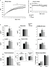
Figure 1
Figure 1
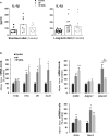
Metabolic status of male rats fed high-fat diets. (A) Body mass and caloric intake of male Wistar rats fed control (8.5% kcal from fat) or high-fat (45% kcal from fat) diets supplemented (HF-DHA) or not (HF) with 5% DHA. (B) Fasting plasma levels of insulin, leptin, glucose, triglycerides and cholesterol (total, free and esterified forms) and body composition (% fat and lean mass) were measured after 11 weeks of diets. The body composition was measured with the Echo MRI™ system. HOMA-IR and TyG indexes were calculated as a reflect of insulin resistance. Data are expressed as means ± SEM (n= 12 rats) and were analyzed with one-way ANOVA followed by Tukey’s multiple comparison test. *P ≤ 0.05; **P ≤ 0.01; ***P ≤ 0.001 vs control. aP ≤ 0.05; bP ≤ 0.01; cP ≤ 0.001 between HF and HF-DHA.
Figure 2
Figure 2
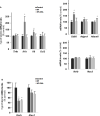
Characterization of peripheral inflammation in rats after 20 weeks of high-fat diets. (A) Circulating levels of the pro-inflammatory cytokine interleukin-1β (IL-1β) in rats fed control, HF or HF-DHA diets for 4 and 20 weeks. Levels of IL-1β were assayed by ELISA as described in Materials and Methods. The bar height reflects the mean and symbols reflect individual animals. (B) Transcript levels of different inflammatory markers in sub-cutaneous adipose tissue after 20 weeks of control or HF diets. Transcript levels were determined by real time PCR, relative levels of genes were normalized to the one of Cyclophilin and expressed as % of the control group. Data are means ± SEM from 11-12 (A) or 8 (B) rats and were analyzed by non-parametric one-way ANOVA (Kruskal-Wallis test) followed by Dunnett’s multiple comparison test. *P ≤ 0.05; **P ≤ 0.01; ***P ≤ 0.001 vs control. bP ≤ 0.01 between HF and HF-DHA.
Figure 3
Figure 3
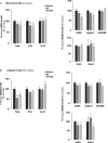
Hypothalamic inflammation and levels of GnRH and Kiss-1 transcripts in rats after 4 weeks of high-fat diets. (A) Transcript levels of pro-inflammatory cytokines (Tnfa, Il1b, Il6, Ccl2), inflammatory-related (Ikkb and Nos2) and macrophage (Cd38, Adgre1, Adam8) genes in hypothalamus of rats fed control, HF or HF-DHA diets for 4 weeks. (B) Hypothalamic transcript levels of Gnrh and Kiss 1 in control and in rats fed short-term diet. Transcript levels were determined and expressed as indicated in Figure 2B. Data are means ± SEM (n= 6-8 rats) and were analyzed with Kruskal-Wallis test followed by Dunnett’s multiple comparison test. *P ≤ 0.05 vs control group.
Figure 4
Figure 4
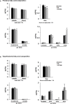
Pituitary inflammation in rats after short-term or long-term high-fat diets. Transcript levels of pro-inflammatory cytokines (Tnfa, Il1b, Il6, Ccl2), inflammation-related (Ikkb and Nos2) and macrophage (Cd38, Adgre1, Adam8) genes in pituitaries of rats fed control, HF or HF-DHA diets for 4 (A) or 20 weeks (B). Transcript levels were determined and expressed as indicated in Figure 2B. Data are expressed as means ± SEM (n= 6-8 rats) and were analyzed with Kruskal-Wallis test followed by Dunnett’s multiple comparison test. *P ≤ 0.05; **P ≤ 0.01; ***P ≤ 0.001 vs respective control.
Figure 5
Figure 5
Fatty acid composition in anterior pituitaries and hypothalamus of rats fed short-term high-fat diets. The concentration of saturated FA (palmitic and stearic acids), monounsaturated FA (oleic acid), ω6 PUFA (linoleic, arachidonic acids) and ω3 PUFA (linolenic acid, eicosapentaenoic acid (EPA), docosapentaenoic acid (DPA) and docosahexaenoic acid (DHA) were determined by GCMS as described in Materials and Methods in pituitary (A) and hypothalamus (B) of rats fed control or high-fat diets for 4 weeks. Results are expressed in μg/mg of tissue and are the mean ± SEM of 5 rats in each group. *P ≤ 0.05; **P ≤ 0.01; ***P ≤ 0.001 compared with control group.; aP ≤ 0.05; cP ≤ 0.001 between HF and HF-DHA groups.
Figure 6
Figure 6
Characterization of pituitary gonadotrope function in rats after short-term or long-term high-fat diets. (A) Transcript levels of gonadotropin subunits, Lhb, Fshb and Cga, were measured in pituitaries of rats fed control, HF or HF-DHA diets for 4 or 20 weeks. Transcript levels were determined and expressed as indicated in Figure 2. (B) The levels of the two circulating gonadotropins, LH and FSH, were simultaneously measured in sera using Luminex technology after 4 and 20 weeks of diet. The bar height reflects the mean. (C) Transcript levels of Gnrhr in control rats or rats fed high-fat diets for 4 and 20 weeks. (D) Pituitary GnRH receptivity in control rats or rats fed high-fat diets for 15 weeks. The natural agonist GnRH (0.5 μg/rat; Sigma LHRH, L 7134, St-Quentin Fallavier, France) was subcutaneously injected and tail blood was collected 0, 5, 15, 30 and 60 min later for LH level determination. The AUC (Area Under the Curve) was calculated for the three groups of rats and no statistical difference was observed. Data are from 6-8 rats in panels A, C and D and from 7-8 (short-term diets) or 11-12 (long-term diets) rats in panel (B) Data are expressed as means ± SEM and analyzed with one-way ANOVA followed by Tukey’s multiple comparison test. *P ≤ 0.05; **P ≤ 0.01; ***P ≤ 0.001 compared to control group.
Figure 7
Figure 7
Circulating levels of non-gonadotrope pituitary hormones rats after short-term or long-term high-fat diets. Serum levels of the pituitary hormones, ACTH, GH, PRL and TSH, were measured after 4 and 20 weeks of diet as described in Materials and Methods. The bar height reflects the mean. Data are expressed as means ± SEM (n= 6-8 and n=10-12 rats for short- and long-term diets, respectively) and were analyzed with Kruskal-Wallis test followed by Dunnett’s multiple comparison test. *P ≤ 0.05 vs control. aP ≤ 0.05 between HF and HF-DHA.
Figure 8
Figure 8
Circulating inhibin B and sex steroid levels in rats after short-term or long-term high-fat diets. Serum levels of inhibin B (A) and sex steroids, testosterone and estradiol (B) were determined after 4 and 20 weeks of diet, as described in Materials and Methods. The bar height reflects the mean. Data are expressed as means ± SEM from 7-8 and 10-12 rats for short- and long-term diets, respectively and were analyzed by one-way ANOVA followed by non-parametric one-way ANOVA (Kruskal-Wallis test) followed by Dunnett’s multiple comparison test. *P ≤ 0.05; **P ≤ 0.01 vs control.

References

    1. Counis R, Laverriere JN, Garrel G, Bleux C, Cohen-Tannoudji J, Lerrant Y, et al. . Gonadotropin-Releasing Hormone and the Control of Gonadotrope Function. Reprod Nutr Dev (2005) 45(3):243–54. doi: 10.1051/rnd:2005017 - DOI - PubMed
    1. Bliss SP, Navratil AM, Xie JJ, Roberson MS. GnRH Signaling, the Gonadotrope and Endocrine Control of Fertility. Front Neuroendocrinol (2010) 31(3):322–40. doi: 10.1016/j.yfrne.2010.04.002 - DOI - PMC - PubMed
    1. Adashi EY, Hsueh AJW, Yen SSC. Insulin Enhancement of Luteinizing-Hormone and Follicle-Stimulating-Hormone Release by Cultured Pituitary-Cells. Endocrinology (1981) 108(4):1441–9. doi: 10.1210/endo-108-4-1441 - DOI - PubMed
    1. Sone M, Osamura RY. Leptin and the Pituitary. Pituitary (2001) 4(1-2):15–23. doi: 10.1023/A:1012978525492 - DOI - PubMed
    1. Fernandez-Fernandez R, Tena-Sempere M, Navarro VM, Barreiro ML, Castellano JM, Aguilar E, et al. . Effects of Ghrelin Upon Gonadotropin-Releasing Hormone and Gonadotropin Secretion in Adult Female Rats: In Vivo and In Vitro Studies. Neuroendocrinology (2005) 82(5-6):245–55. doi: 10.1159/000092753 - DOI - PubMed

Publication types