. 2022 Apr 14:13:846226.
doi: 10.3389/fendo.2022.846226. eCollection 2022.

Nuclear Progesterone Receptor Expressed by the Cortical Thymic Epithelial Cells Dictates Thymus Involution in Murine Pregnancy

Affiliations

Nuclear Progesterone Receptor Expressed by the Cortical Thymic Epithelial Cells Dictates Thymus Involution in Murine Pregnancy

Soo Hyun Ahn et al. Front Endocrinol (Lausanne). .

Erratum in

Abstract

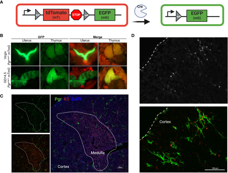
Progesterone is a gonadal pro-gestational hormone that is absolutely necessary for the success of pregnancy. Most notable actions of progesterone are observed in the female reproductive organs, the uterus and the ovary. Acting through the nuclear progesterone receptor (PGR), progesterone prepares the endometrium for implantation of the embryo. Interestingly, the maternal thymus also is a known expressor of Pgr; its absence is associated with murine pregnancy complications. However, the localization of its expression and its functional importance were not known. Here, we used a transgenic dual fluorescent reporter mouse model and genetic deletion of Pgr in Foxn1+ thymic epithelial cells (TEC) to demonstrate TEC-specific Pgr expression in pregnancy, especially in the cortex where thymocyte maturation occurs. Using our TEC-specific Pgr deletion mouse model, we demonstrate that TEC-specific Pgr is necessary for pregnancy-induced thymic involution in pregnancy. Our investigation reveals that PGR expression is upregulated in the cortical thymic epithelial cells during pregnancy, and that PGR expression is important for thymic involution during murine pregnancy.

Keywords: fertility; involution; pregnancy; progesterone receptor (PGR); thymus.


Conflict of interest statement

The authors declare that the research was conducted in the absence of any commercial or financial relationships that could be construed as a potential conflict of interest. The reviewer S-PW declared a past collaboration with two of the authors JL and J-WJ to the handling editor.

Figures

Figure 1
Figure 1
Thymus involution in pregnancy. (A) Comparison of gross anatomy of female murine thymus in non pregnant (NP) and GD18.5. (B) thymus weight throughout pregnancy. (C) Total cell number from thymus throughout pregnancy. (D) Immunofluorescence staining of NP and GD18.5 thymus for cytokeratin 5 (K5) and cytokeratin 8 (K8) denoting medulla and cortex, respectively, to illustrate the changes in the volume occupied by each compartment in pregnancy. scale bar = 500μm. (E) Comparison of area coverage by medulla (red) to DAPI (entire surface area) in NP and GD15.5 thymus using IMARIS surface rendering function. scale bar = 1000μm. Statistical analysis was performed by non-parametric Kruskal-Wallis test followed by Dunn post hoc test. *p < 0.05, Each dot represents one mouse.
Figure 2
Figure 2
Nuclear Progesterone Receptor expression increases in the thymus in pregnancy. (A) RT-qPCR for Esr1, Esr2, Pgr, Pgrmc1, and Pgrmc2 between NP (n=3) and P (GD16.5, n=5) thymus. Each dot represents one mouse. (B) % PR positive nuclei calculated from field of DAPI from NP (n=4), GD6.5 (n=3), GD14.5 (n=6), GD16.5 (n=3), and GD18.5 (n=4) thymus; see Supplementary Figure 1 for representative images. *p < 0.05, ***p < 0.0001; one-way ANOVA with Dunnett’s post-hoc test with comparisons to NP controls. Each dot represents one mouse. (C) Immunofluorescence staining of GD14.5 thymus with antibodies specific to cytokeratin 8 (K8) and nuclear PGR. C, cortex; M, medulla, Scale bar = 100μm, 200x magnification.
Figure 3
Figure 3
Pgr reporter system identifies PGR + cells in the cortex of the thymus in pregnancy. (A) Explanation of Cre/Rosa26mTmG reporter system. In the absence of Cre recombinase activity, all cells remain mT+ (red). Upon expression of Pgr gene, Cre recombinase generation mediates the excision of the mT cassette. Post recombination, the cell membrane of all PGR+ cells will become GFP+. (B) Macroscopic visualization of GFP expression in the thymus at GD14.5 compared to virgin control. (C) Partially stitched Pgrcre/+;Rosa26mTmG/mTmG thymus at GD14.5 to demonstrate GFP+ expression in the cortex of the thymus. K5 denotes medulla. DAPI = nuclei, PGR = GFP+ cells. The dotted lines outline the medulla. Magnification: 200x, Scale bar = 100μm. (D) Pgrcre/+;Rosa26mTmG/mTmG thymus at GD14.5 stained with anti-PGR primary antibody and counter stained with anti-rabbit secondary antibody (Alexa Fluor 405), pseudo-colored red to provide contrast to GFP+ cells. Dotted line indicates the capsule of the thymus. Magnification: 200x, Scale bar = 100μm.
Figure 4
Figure 4
Conditional deletion of Pgr gene from the Foxn1+ thymic epithelial cells prevents thymic involution in pregnancy. (A) Nuclear progesterone receptor (PGR) colocalizes with Foxn1+ Thymic epithelial cells (TECs) at GD6.5. Alexa flour 405 was used as a secondary antibody. PGR is pseudo-colored red to provide contrast to the GFP+. 400x magnification, scale bar = 50μm. (B) Immunofluorescence staining to detect PGR protein confirms the conditional deletion of Pgr in the thymus at GD16.5 of Pgrd/d females. PGR expression is intact in the NP female uterus of both Pgrf/f controls and Pgrd/d animals. Magnification at 200x. Scale bar = 100μm, (C) Thymic weight (Top) and cellularity (Bottom) in NP and GD16.5 C57BL6WT, Pgrf/f /Pgrf/+ (FLFL/FLWT) or Pgrd/d (FLKO) females. Red dots = GD14.5. (D) Thymic weight (Top) and cellularity (Bottom) in C57BL6WT, Pgrf/f /Pgrf/+ (FLFL/FLWT) or Pgrd/d (FLKO) between NP and pregnant (GD14.5 - GD16.5) females. Each dot represents one mouse. Statistical analysis was performed using non-parametric Wilcoxon test (C) or Kruskal-Wallis test followed by Dunn’s test for post-hoc assessment with Bonferroni correction (D). P<0.05 deemed significant. **p < 0.001, ***p < 0.0005, ****p < 0.00001, ns, non-significant.
Figure 5
Figure 5
Thymocyte developmental process is undisrupted in the thymus of Pgrd/d in pregnancy. (A) Gating strategy of the murine thymus for CD4SP, CD8SP, DN, and DP. (B) % CD4 single positive (SP), CD8SP and DN in WT, FLFL/FLWT and FLKO between NP and GD14.5-GD16.5 of pregnancy. (C) % DP population in WT, FLFL/FLWT and FLKO between NP and GD14.5-GD16.5 of pregnancy. Student’s t-test, *p < 0.05, ns, not significant. Each dot represents one mouse.
Figure 6
Figure 6
Pgrd/d thymus show unchanged expression of Fezf2, involved in self-antigen expression and decreased expression of Tnfrsf11a (RANK), involved in mTEC maintenance. Comparison of gene expression in the thymus of Pgrf/+ or Pgrf/f (FLWT/FLFL) and Pgrd/d (FLKO) females between non-pregnant (NP) and GD16.5 of pregnancy. Delta CT was calculated using Gapdh as housekeeping gene. Delta CT of each gene from C57BL/6J WT NP thymus samples were used to calculate delta-delta CT, then the values were Log2 transformed to calculate Log2FoldChange (Log2FC). *p < 0.05, **p < 0.01, ***p < 0.001. Each dot represents one mouse.
Figure 7
Figure 7
Assessment of long-term fertility outcome in FLFL/FLWT (Pgrf/f or Pgrf/+ ) and FLKO (Pgrd/d ) in allogeneic and syngeneic mating strategy. (A) Individual litter size from female of FLFL/FLWT and FLKO mated with either allogeneic (Balb/c) or syngeneic (C57) male. (B) Weight of male or female 3 week old weanlings born to FLFL/FLWT and FLKO from allogeneic or syngeneic mating. **p < 0.01, ****p < 0.0001, ns, not-significant. (C) Cumulative numbers of pups across three allogeneic pregnancies in controls (Flox-1 to Flox- 3) and FLKO animals (KO-1 to KO-3).

References

    1. Petrie HT, Zúñiga-Pflücker JC. Zoned Out: Functional Mapping of Stromal Signaling Microenvironments in the Thymus. Annu Rev Immunol (2007) 25:649–79. doi: 10.1146/annurev.immunol.23.021704.115715 - DOI - PubMed
    1. Nehls M, Pfeifer D, Schorpp M, Hedrich H, Boehm T. New Member of the Winged-Helix Protein Family Disrupted in Mouse and Rat Nude Mutations. Nature (1994) 372:103–7. doi: 10.1038/372103a0 - DOI - PubMed
    1. Moore TA, von Freeden-Jeffry U, Murray R, Zlotnik A. Inhibition of Gamma Delta T Cell Development and Early Thymocyte Maturation in IL-7 -/- Mice. J Immunol (1996) 157:2366–73. - PubMed
    1. Clarke AG, Kendall MD. The Thymus in Pregnancy: The Interplay of Neural, Endocrine and Immune Influences. Immunol Today (1994) 15:545–51. doi: 10.1016/0167-5699(94)90212-7 - DOI - PubMed
    1. Tibbetts TA, DeMayo F, Rich S, Conneely OM, O’Malley BW. Progesterone Receptors in the Thymus are Required for Thymic Involution During Pregnancy and for Normal Fertility. Proc Natl Acad Sci USA (1999) 96:12021–6. doi: 10.1073/pnas.96.21.12021 - DOI - PMC - PubMed