. 2020 Mar 10;10(17):10118-10128.
doi: 10.1039/c9ra09710d. eCollection 2020 Mar 6.

Preparation and biological properties of silk fibroin/nano-hydroxyapatite/graphene oxide scaffolds with an oriented channel-like structure

Affiliations

Preparation and biological properties of silk fibroin/nano-hydroxyapatite/graphene oxide scaffolds with an oriented channel-like structure

Lu Wang et al. RSC Adv. .

Abstract

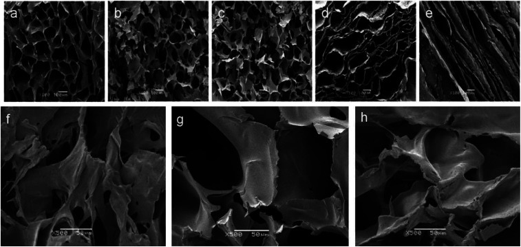
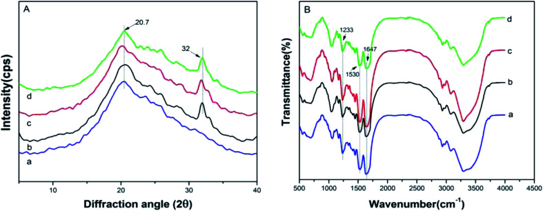
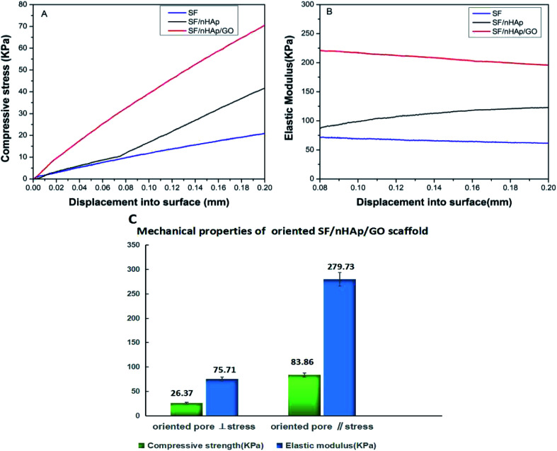
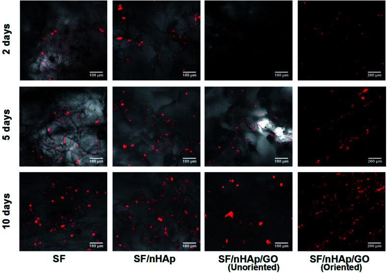
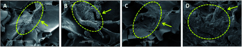
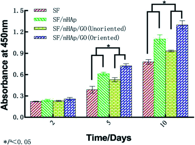
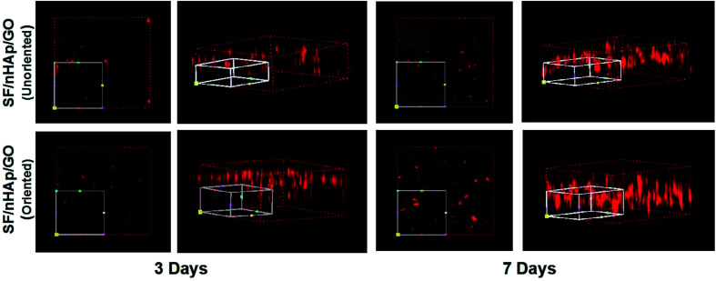
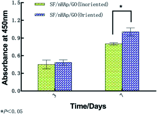
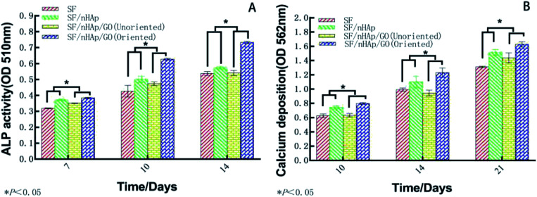
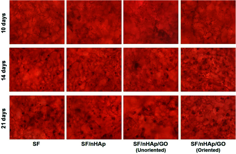
Constructing an ideal bone tissue engineering scaffold has been one of the research hotspots in the biomedical field. Silk fibroin (SF), nano-hydroxyapatite (nHAp) and graphene oxide (GO) are excellent biomaterials, and have been studied and explored extensively. To better mimic natural bone, we fabricated a SF/nHAp/GO hybrid scaffold with an oriented channel-like structure by using directional temperature field freezing technology. A comparative analysis was carried out for the SF, SF/nHAp, unoriented SF/nHAp/GO and oriented SF/nHAp/GO scaffolds. The physical and chemical properties were studied by scanning electron microscopy, X-ray diffraction, Fourier transform infrared spectroscopy and universal mechanical testing. The data showed that the oriented channel-like SF/nHAp/GO porous scaffold expressed high interconnectivity, suitable pore diameter and porosity and anisotropic mechanical properties. Cytocompatibility tests indicated that the oriented channel-like SF/nHAp/GO porous scaffold was more favorable for stimulating bone marrow mesenchymal stem cells (BMSCs) adhesion and proliferation. Additionally, human umbilical vein endothelial cells (HUVECs) grew unimpeded along the channel, indicating it had advantages for vascularization. For further testing in vitro, osteogenic induction was carried out on BMSCs inoculated on the above scaffolds, and then alkaline phosphatase (ALP) activity was tested and cell mineralization was observed. The results indicated that the oriented channel-like SF/nHAp/GO porous scaffold was more conducive to osteogenic differentiation of BMSCs. Hence, the material may prove to be a promising scaffold for bone tissue engineering.


Conflict of interest statement

The authors declare no conflict of interest.

Figures

Fig. 1
Fig. 1. SEM images of unoriented and oriented porous scaffolds. (a) SF (b) SF/nHAp (c) unoriented SF/nHAp/GO (d) cross-section of oriented SF/nHAp/GO. (e) Longitudinal-section of oriented SF/nHAp/GO (×100) (f) SF (g) SF/nHAp (h) unoriented SF/nHAp/GO (×500).
Fig. 2
Fig. 2. XRD (A) and FT-IR (B) of SF, SF/nHAp, unoriented SF/nHAp/GO and oriented SF/nHAp/GO scaffolds. (a) SF (b) SF/nHAp (c) unoriented SF/nHAp/GO (d) oriented SF/nHAp/GO.
Fig. 3
Fig. 3. Compressive stress (A) and elastic modulus (B) of SF, SF/nHAp, unoriented. SF/nHAp/GO scaffolds. Mechanical properties (C) of oriented SF/nHAp/GO scaffold.
Fig. 4
Fig. 4. LSCM images to show BMSCs cultured on different scaffolds for 2, 5 and 10 days.
Fig. 5
Fig. 5. SEM images of BMSCs cultured on different scaffolds for 10 days. (The dotted areas pointed by the arrow are cell masses surrounded by matrix.) (A) SF (B) SF/nHAp (C) unoriented SF/nHAp/GO (D) Oriented SF/nHAp/GO.
Fig. 6
Fig. 6. OD Values of BMSCs cultured on the different scaffolds for 2, 5 and 10 days.
Fig. 7
Fig. 7. 3-Dimension LSCM images to show HUVECs cultured on the unoriented and oriented SF/nHAp/GO porous scaffolds for 3 and 7 days.
Fig. 8
Fig. 8. OD Values of HUVECs cultured on the unoriented and oriented SF/nHAp/GO. Porous scaffolds for 3 and 7 days.
Fig. 9
Fig. 9. Alkaline phosphatase in BMSCs cultured on the different scaffolds for 7, 10 and 14 days (A). Calcium deposition in the different scaffolds for 10, 14 and 21 days (B).
Fig. 10
Fig. 10. Alizarin red staining of BMSCs growing on different scaffolds for 14 and 21 days.

References

    1. Lu Q. Hu X. Wang X. Q. Kluge J. A. Lu S. Z. Kaplan D. L. Acta Biomater. 2010;6(4):1380–1387. doi: 10.1016/j.actbio.2009.10.041. - DOI - PMC - PubMed
    1. Nazarov R. Jin H. J. Kaplan D. L. Biomacromolecules. 2004;5(3):718–726. doi: 10.1021/bm034327e. - DOI - PubMed
    1. Kim U. J. Park J. Li C. M. Jin H. J. Valluzzi R. Kaplan D. L. Biomacromolecules. 2004;5(3):786–792. doi: 10.1021/bm0345460. - DOI - PubMed
    1. Schneider A. Wang X. Y. Kaplan D. L. Garlick J. A. Egles C. Acta Biomater. 2009;5(7):2570–2578. doi: 10.1016/j.actbio.2008.12.013. - DOI - PMC - PubMed
    1. Meinel L. Fajardo R. Hofmann S. Kaplan D. L. Bone. 2005;37(5):688–698. doi: 10.1016/j.bone.2005.06.010. - DOI - PubMed