. 2021 Oct 5:37:91-106.
doi: 10.1016/j.jare.2021.10.001. eCollection 2022 Mar.

Hypoxia inducible lncRNA-CBSLR modulates ferroptosis through m6A-YTHDF2-dependent modulation of CBS in gastric cancer

Affiliations

Hypoxia inducible lncRNA-CBSLR modulates ferroptosis through m6A-YTHDF2-dependent modulation of CBS in gastric cancer

Hui Yang et al. J Adv Res. .

Abstract

Introduction: Tumors are usually refractory to anti-cancer therapeutics under hypoxic conditions. However, the underlying molecular mechanism remains to be elucidated.

Objectives: Our study intended to identify hypoxia inducible lncRNAs and their biological function in gastric cancer (GC).

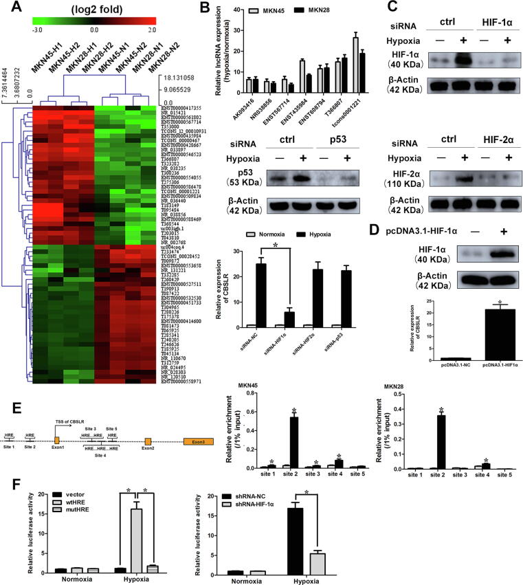
Methods: Differentially expressed lncRNAs were determined by microarray analysis between GC cells exposed to hypoxia (1% O2) and normoxia (21% O2) for 24 h. The expression level of CBSLR was manipulated in several GC cell lines to perform molecular and biological analyses both in vitro and in vivo.

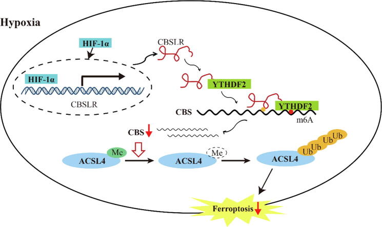
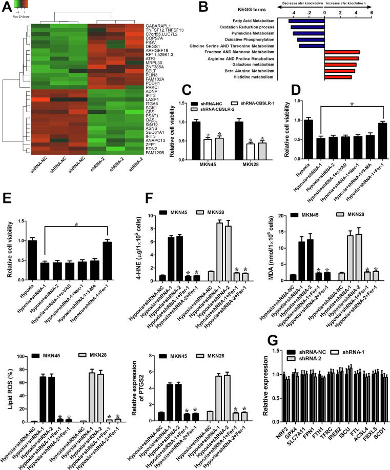
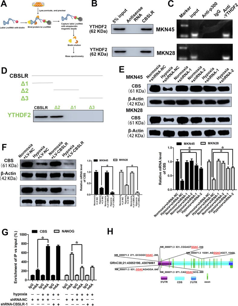
Results: We identified a hypoxia-induced lncRNA-CBSLR that protected GC cells from ferroptosis, leading to chem-resistance. Mechanically, CBSLR interacted with YTHDF2 to form a CBSLR/YTHDF2/CBS signaling axis that decreased the stability of CBS mRNA by enhancing the binding of YTHDF2 with the m6A-modified coding sequence (CDS) of CBS mRNA. Furthermore, under decreased CBS levels, the methylation of the ACSL4 protein was reduced, leading to protein polyubiquitination and degradation of ACSL4. This, in turn, decreased the pro-ferroptosis phosphatidylethanolamine (PE) (18:0/20:4) and PE (18:0/22:4) content and contributed to ferroptosis resistance. Notably, CBSLR is upregulated, whereas CBS is downregulated in GC tissues compared to matched normal tissues; and GC patients with high CBSLR/low CBS levels have a worse clinical outcome and a poorer response to chemotherapy.

Conclusion: Our study reveals a novel mechanism in how HIF1α/CBSLR modulates ferroptosis/chemoresistance in GC, illuminating potential therapeutic targets for refractory hypoxic tumors.

Keywords: 4-HNE, 4-hydroxynonenal; AJCC, American Joint Committee on Cancer; CDS, coding sequence; CHIP, chromatin immunoprecipitation; Chemoresistance; DZNeP, 3-deazaneplanocin A; FBS, fetal bovine serum; Ferroptosis; GC, gastric cancer; GEO, Gene Expression Omnibus; Gastric cancer; HE, Hematoxylin and eosin; HREs, hypoxia response elements; Hypoxia; IHC, immunohistochemical; MDA, malondialdehyde; RIP, RNA immunoprecipitation; TCGA, the cancer genome atlas; TNM, tumor-node-metastasis staging system; YTHDF2, YTH domain family protein 2; lncRNA; lncRNA, long noncoding RNA; qPCR, quantitative real-time PCR.


Conflict of interest statement

The authors declare that they have no known competing financial interests or personal relationships that could have appeared to influence the work reported in this paper.

Figures

None
Graphical abstract
Fig. 1
Fig. 1
tcons1221 expression is induced upon hypoxia. (A) hierarchical clustering analysis of the top 60 lncRNAs (30 upregulated and 30 downregulated) that were differentially expressed between hypoxia-treated (1% O2, 24 h) and normoxia-treated (21% O2) GC cells (MKN45 and MKN28 cell lines). (B) The expression profiling of seven cancer-associated lncRNAs in GC cells under hypoxia (1% O2, 24 h). (C) MKN45 cells were transfected with siRNA-NC, siRNA-HIF-1α, siRNA-HIF-2α, or siRNA-p53. Twenty-four hours after transfection, cells were cultured under normoxic or hypoxic conditions for 24 h. The expression levels of target genes were determined by western blot analysis. The levels of CBSLR expression were determined by qRT-PCR. (D) MKN45 cells were transfected with pcDNA3.1-HIF-1α. Twenty-four hours after transfection, the expression level of HIF-1α was determined by western blot analysis. The levels of CBSLR expression were examined by qRT-PCR. (E) Schematic illustration of HIF-1α responsive element (HRE) in CBSLR locus. MKN45 and MKN28 cells were cultured under normoxia or hypoxic conditions (1% O2) for 24 h, then, a CHIP assay was employed to examine the binding of HIF-1α to each HRE. (F) (Upper) MKN45 cells were cotransfected with Renilla luciferase plasmid and the indicated reporter constructs. (Lower) MKN45 cells expressing shRNA-NC or shRNA-HIF-1a were cotransfected with Renilla luciferase plasmid and reporter constructs containing the sequence around the second HRE site. Twenty-four hours after transfection, cells were cultured under normoxic or hypoxic conditions (1% O2) for 24 h. Luciferase activity was then determined and normalized to Renilla luciferase activity. Results were expressed as mean ± S.D. (n = 3).
Fig. 2
Fig. 2
CBSLR silencing induced ferroptosis and rescued chemoresistance in GC. (A) The heat map demonstrates the top 32 differentially expressed genes (16 upregulated and 16 downregulated) between shRNA-CBSLR and shRNA-NC MKN45 cells cultured under hypoxic conditions for 24 h, with three repeats. (B) Top five GSEA pathways identified by KEGG analysis (false discovery rate of < 0.1) for the differentially expressed genes in (A). The pathways are ranked by normalized enrichment score (NES). (C) Proliferative activity of shRNA-NC and shRNA-CBSLR MKN45/MKN28 cells grown for 24 h of hypoxia and assessed by CellTiter Glo assay. (D, E) Inhibitors of apoptosis (20 μM z-VAD), autophagy (0.5 mM 3-MA), necroptosis (10 μM Necrostatin-1), and ferroptosis (1 μM Fer-1) were added to the shRNA-NC or shRNA-CBSLR MKN45 (D) or MKN28 (E) cells. The cells were cultured under hypoxia (1% O2 for 24 h). Twenty-four hours later, the Celltiter Glo assay was utilized to determine the cell viability. (F) Inhibitors of ferroptosis (1 μM Fer-1) were added to the shRNA-NC or shRNA-CBSLR MKN45 or MKN28 cells. The cells were cultured under hypoxia (1% O2 for 24 h). Twenty-four hours later, MDA and 4-HNE levels were examined in different groups, with three repeats. The levels of lipid peroxidation and PTGS2 were detected using BODIPY staining and qPCR, respectively, with three repeats. *, p < 0.05, significantly different from shRNA-CBSLR transfected GC cells exposed to hypoxia (1% O2 for 24 h). (G) MKN45 cells transfected with shRNA-NC or shRNA-CBSLR-1,2 were cultured under hypoxic conditions (1% O2 for 24 h). Twenty-four hours later, gene levels were detected through qRT-PCR. Results were expressed as mean ± S.D. (n = 3).
Fig. 3
Fig. 3
CBSLR physically interacts with YTHDF2 and modulates CBS level. (A) Schematic overview of in vitro RNA Antisense Purification and used for identification of proteins interacting with CBSLR. (B) RNA pull-down assay was performed to confirm the association between YTHDF2 and CBSLR. (C) RIP experiments shows the binding of CBSLR with YTHDF2. (D) RNA pull-down using sequentially deleted CBSLR fragments demonstrates the binding segment of CBSLR with YTHDF2. (E) shRNA-NC or shRNA-CBSLR MKN45 or MKN28 cells were cultured under normoxic or hypoxic conditions for 24 h. The mRNA and protein levels of CBS were determined using qRT-PCR or western blot analysis. (F) MKN45, and MKN28 cells stably infected with lentiviruses encoding lncRNA-CBSLR or NC were cultured under normoxic or hypoxic conditions for 24 h. The mRNA and protein levels of CBS were determined using qRT-PCR or western blot analysis. (G) The shRNA-NC and shRNA-CBSLR-1-MKN45 cells were cultured under normal or hypoxic conditions (24 h). m6A RIP-qPCR analysis of CBS or NANOG (positive control) mRNA was performed. (H) Schematic illustrations of m6A motifs positions of CBS mRNA.
Fig. 4
Fig. 4
m6A regulates the mRNA stability of CBS. (A) The shRNA-NC and shRNA-METTL3 MKN45 and MKN28 cells were cultured under hypoxic conditions for 24 h. m6A RIP-qPCR analysis of CBS mRNA was performed. (B) (Upper) The shRNA-NC or shRNA-METTL3-MKN45 cells were cultured under hypoxic conditions for 24 h. (Lower) MKN45 cells were pretreated with DZNeP (10 μM) for 1 h and then cultured under hypoxic conditions (24 h). m6A-RIP-qPCR analysis was performed to evaluate the m6A in 5′UTR or CDS of CBS with fragmented RNA. (C) (Left) MKN45 and MKN28 cells pretreated with DZNeP (10 μM) or DMSO for 1 h and then cultured normoxic or hypoxic conditions for 24 h. (Right) LV-NC or LV-CBSLR-MKN45 and MKN28 cells were pretreated with DZNeP (10 μM) or DMSO and then cultured under hypoxic conditions (24 h). The protein level of CBS was determined by western blot analysis. (D) Schematic representation of mutated 5′UTR. shRNA-NC or shRNA-METTL3-MKN45 cells were cotransfected with pGL3-CBS-5′UTR-WT or pGL3-CBS-5′UTR-Mut reporter. Twenty-four hours later, the cells were cultured under hypoxic conditions for 24 h. The protein, mRNA and translation efficiency were determined. (E) Schematic illustrations of mutation in m6A motif in CDS of CBS. shRNA-NC or shRNA-METTL3-MKN45 cells were cotransfected with pmirGLO-CBS-CDS-WT or pmirGLO-CBS-CDS-Mut1 reporter. Twenty-four hours later, the cells were cultured under hypoxic conditions for 24 h. The protein and mRNA were determined.
Fig. 5
Fig. 5
CBSLR/YDHDF2/CBS ternary complex promotes degradation of CBS mRNA. (A) pcDNA3.1-CBS-CDS-WT or pcDNA3.1-CBS-CDS-Mut was cotransfected into shRNA-NC or shRNA- METTL3-MKN45 cells. After 24 h, the cells were cultured under hypoxic conditions for 24 h. Cell lysates were subjected to western blot analysis. (B) pcDNA3.1-CBS-CDS-WT or pcDNA3.1-CBS-CDS-Mut was transfected into shRNA-NC or shRNA-METTL3-MKN45 cells. After 24 h, the cells were cultured under hypoxic conditions for 20 h and then further treated with Act-D (0.5 mg/mL) for the indicated times. The mRNA level of CBS was examined by qRT-PCR. (C) The MKN45 cells were cultured under normoxic or hypoxic conditions for 24 h. RIP experiments were performed using the YTHDF2 antibody for immunoprecipitation and qRT-PCR was utilized to detect CBSLR. (D) shRNA-NC or shRNA-METTL3- MKN45 or MKN28 cells were transfected with pcDNA3.1-NC or pcDNA3.1-YTHDF2. Twenty-four hours later, the cells were cultured under hypoxia for 24 h. The protein level of CBS was evaluated by western blot analysis. (E, F) Schematic diagram of CBS WT (CBS-CDS-WT-luc) and CBS mutant (CBS-CDS-MUT-luc) reporters (E). The MKN45 cells were transfected with pmirGLO-CBS-CDS-WT or pmirGLO-CBS-CDS-Mut1 reporter. After 24 h, MKN45 cells were cultured under hypoxia for 24 h. Relative luciferase activity was evaluated. (G, H) shRNA-NC or shRNA-CBSLR-MKN45 cells were cotransfected with pmirGLO-CBS-CDS-WT or pmirGLO-CBS-CDS-Mut1 reporter. MKN45 stably infected with lentiviruses encoding lncRNA-CBSLR or NC were cotransfected with pmirGLO-CBS-CDS-WT or pmirGLO-CBS-CDS-Mut1 reporter. Twenty-four hours later, the cells were cultured under hypoxic conditions for 24 h. Relative luciferase activity and luciferase mRNA expression were determined. (I) RIP assay reveals the relative association of YTHDF2 with CBS mRNA upon CBSLR downregulation or upregulation. (J) shRNA-NC or shRNA-CBSLR-MKN45 cells were transfected with pcDNA3.1-NC or pcDNA3.1-YTHDF2. Twenty-four hours later, the cells were cultured under hypoxic conditions for 20 h in the presence or absence of DZNeP (10 μM). A time course started with treating cells with Act-D (0.5 mg/mL) for the indicated times. The CBS mRNA was examined with qRT-PCR. Data represent mean ± S.D. from three independent experiments.
Fig. 6
Fig. 6
CBSLR/CBS correlated with survival rate and response to chemotherapeutics in GC. (A) Kaplan–Meier curves for overall survival and disease-free survival in the CBSLR-high or CBSLR-low groups. (B) The representative images of negative, weak, moderate and strong CBS expression patterns in GC according to IHC staining (×200). (C) Kaplan–Meier curves for overall survival and disease-free survival in the CBS-high or CBS-low groups. (D) Spearman rank-correlation analysis was performed to the correlation between the ΔCt values (normalized to β-actin) of CBSLR and IHC score of CBS in 109 GC tissues. (E) CBSLR knockdown, CBS knockout or control MKN45 cells were grown as xenografts. DDP treatment started after tumors reached approximately 50 mm3 in size. DDP: 7 mg per kg, once a week for three weeks. (F) Xenograft tumor masses were harvested. Tumor weights were determined. (G) The mRNA level of PTGS2 in the indicated tumor was examined with qRT-PCR. (H) The 4-HNE contents in different groups were determined.
Fig. 7
Fig. 7
CBSLR inhibits ferroptosis by modulating ACSL4 methylation to be polyubiquitinated. (A) shRNA-NC or shRNA-CBSLR-MKN45 cells were cultured under hypoxic conditions for 24 h. Cell lysates were subjected to western blot analysis. (B) siRNA-NC or siRNA-CBS was cotransfected into shRNA-NC or shRNA- CBSLR-MKN45 cells. After 24 h, the cells were cultured under hypoxic conditions (24 h). Anti-dimethyl-ACSL4 and anti-ACSL4 antibodies were used in the western blot analysis. The transcript level of ACSL4 was examined with qRT-PCR. (C) ACSL4 endogenous polyubiquitination ((Ub)n-ACSL4) was observed to be decreased with CBSLR silencing and to be increased with CBS silencing in MKN45 cells under hypoxic conditions (24 h). (D) shRNA-NC, shRNA-CBSLR, siRNA-NC, siRNA-CBS-MKN45 cells were cultured under hypoxic conditions for 18 h. MG132 (10 mM) was treated for 6 h. Cell lysates were analyzed by western blot with anti-dimethyl-ACSL4 and anti-ACSL4 antibodies. (E) Heat map of all major PE species in MKN45 cells with hierarchical clustering of shRNA-NC + siRNA-NC, shRNA-CBSLR + siRNA-NC, shRNA-NC + siRNA-CBS, shRNA-CBSLR + siRNA-CBS. Each PE species was normalized to the corresponding mean value. (F) Schematic diagram showing that HIF-1α induces lncRNA-CBSLR to recruit YTHDF2 protein and CBS mRNA to form CBSLR/ YTHDF2/CBS complex, which in turn decreases CBS mRNA stability in an m6A dependent manner. The decreased CBS expression reduced methylation of ACSL4 protein, thus, the protein is degraded via the ubiquitination-proteasome pathway. It eventually protects GC from ferroptosis under a hypoxic tumor microenvironment.

References

    1. Sung H., Ferlay J., Siegel R.L., Laversanne M., Soerjomataram I., Jemal A., et al. Global Cancer Statistics 2020: GLOBOCAN Estimates of Incidence and Mortality Worldwide for 36 Cancers in 185 Countries. CA Cancer J Clin. 2021;71(3):209–249. - PubMed
    1. Chen W., Zheng R., Baade P.D., Zhang S., Zeng H., Bray F., et al. Cancer statistics in China, 2015. CA Cancer J Clin. 2016;66(2):115–132. - PubMed
    1. Smyth E.C., Nilsson M., Grabsch H.I., van Grieken N.CT., Lordick F. Smyth, Magnus Nilsson, Heike I Grabsch, Nicole Ct van Grieken Florian Lordick. Gastric cancer. Lancet. 2020;396(10251):635–648. - PubMed
    1. de Heer E.C., Jalving M., Harris A.L. HIFs, angiogenesis, and metabolism: elusive enemies in breast cancer. J Clin Invest. 2020;130(10):5074–5087. - PMC - PubMed
    1. Bhandari V., Hoey C., Liu L.Y., Lalonde E., Ray J., Livingstone J., et al. Molecular landmarks of tumor hypoxia across cancer types. Nat Genet. 2019;51(2):308–318. - PubMed

Publication types