. 2022 May 2;132(9):e156774.
doi: 10.1172/JCI156774.

HIF inhibitor 32-134D eradicates murine hepatocellular carcinoma in combination with anti-PD1 therapy

Affiliations

HIF inhibitor 32-134D eradicates murine hepatocellular carcinoma in combination with anti-PD1 therapy

Shaima Salman et al. J Clin Invest. .

Abstract

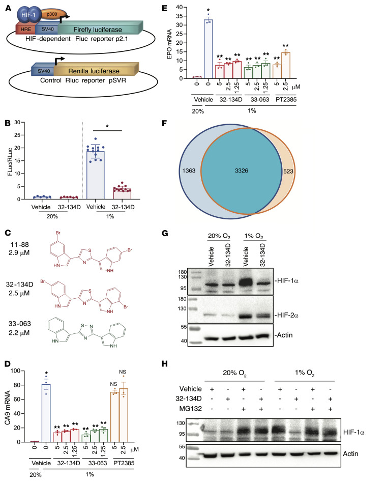
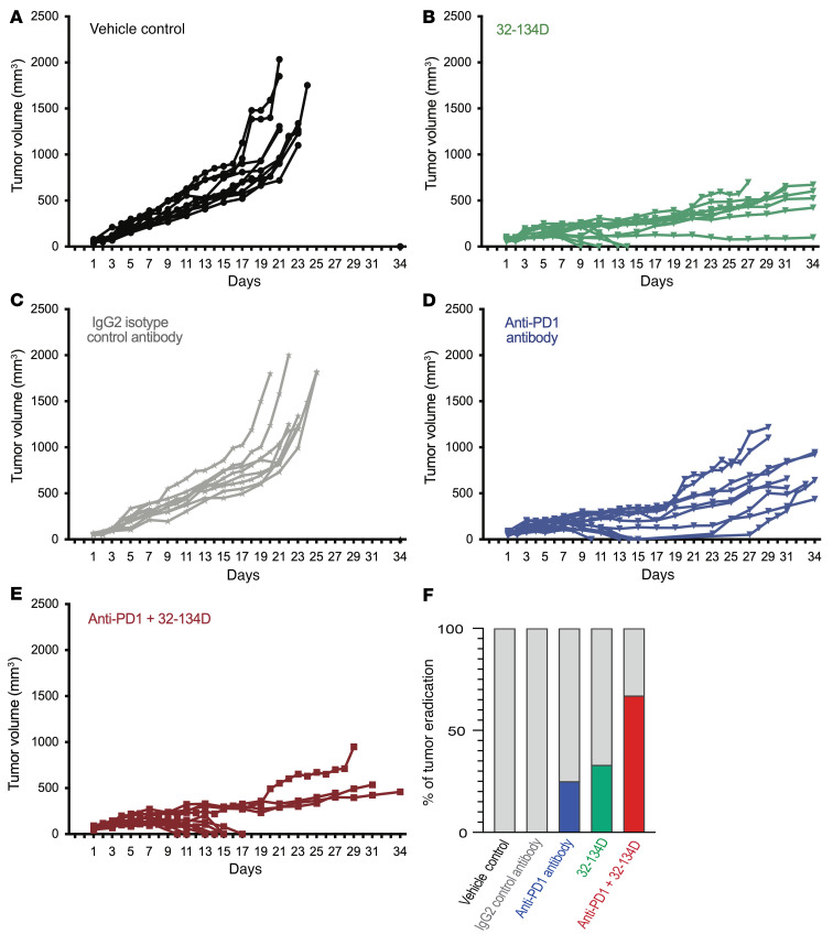
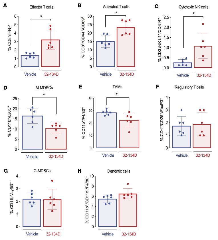
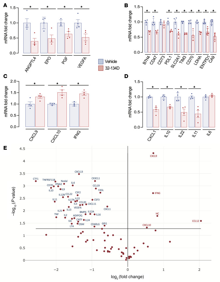
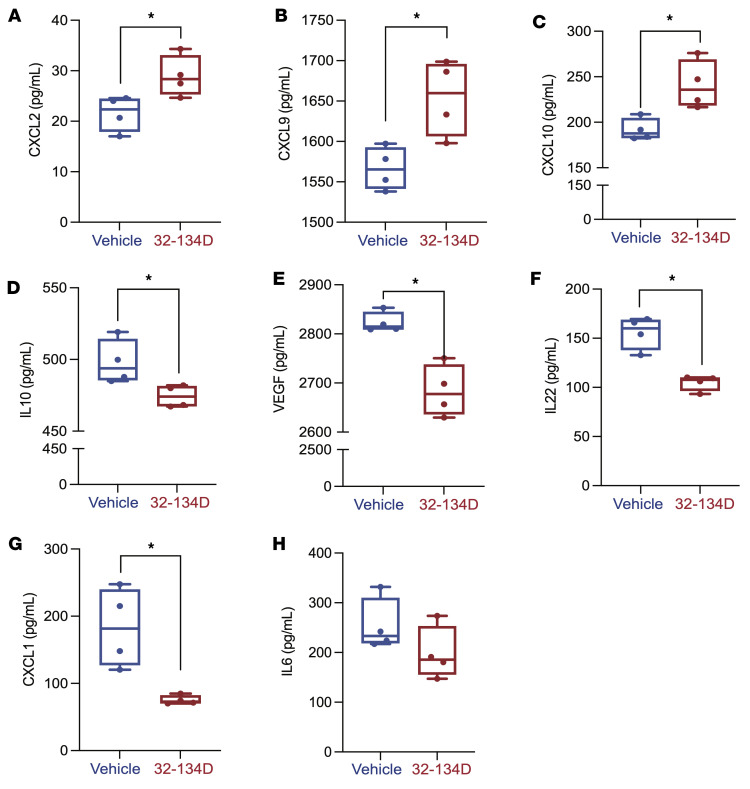
Hepatocellular carcinoma (HCC) is a major cause of cancer mortality worldwide and available therapies, including immunotherapies, are ineffective for many patients. HCC is characterized by intratumoral hypoxia, and increased expression of hypoxia-inducible factor 1α (HIF-1α) in diagnostic biopsies is associated with patient mortality. Here we report the development of 32-134D, a low-molecular-weight compound that effectively inhibits gene expression mediated by HIF-1 and HIF-2 in HCC cells, and blocks human and mouse HCC tumor growth. In immunocompetent mice bearing Hepa1-6 HCC tumors, addition of 32-134D to anti-PD1 therapy increased the rate of tumor eradication from 25% to 67%. Treated mice showed no changes in appearance, behavior, body weight, hemoglobin, or hematocrit. Compound 32-134D altered the expression of a large battery of genes encoding proteins that mediate angiogenesis, glycolytic metabolism, and responses to innate and adaptive immunity. This altered gene expression led to significant changes in the tumor immune microenvironment, including a decreased percentage of tumor-associated macrophages and myeloid-derived suppressor cells, which mediate immune evasion, and an increased percentage of CD8+ T cells and natural killer cells, which mediate antitumor immunity. Taken together, these preclinical findings suggest that combining 32-134D with immune checkpoint blockade may represent a breakthrough therapy for HCC.

Keywords: Cancer immunotherapy; Liver cancer; Oncology; Therapeutics; Transcription.


Conflict of interest statement

Conflict of interest: SS, DJM, YH, and GLS are inventors on provisional patent application US 63/231,216. GLS is a founder of and holds equity in HIF Therapeutics, Inc. This arrangement has been reviewed and approved by the Johns Hopkins University in accordance with its conflict-of-interest policies.

Figures

Figure 1
Figure 1. Identification of HIF inhibitors.
(A) Hep3B-c1 cells were stably transfected with firefly luciferase (FLuc) reporter p2.1, which contains a hypoxia-response element (HRE), and Renilla luciferase (RLuc) reporter pSVR. (B) Hep3B-c1 cells were incubated with vehicle (Veh; 0.1% DMSO; blue bars) or 10 μM 32-134D (red bars) at 20% O2 (n = 6) or 1% O2 (n = 12) for 24 hours. Cell lysates were assayed for Fluc/Rluc activity (mean ± SEM); *P < 0.05 versus vehicle (χ2 test). (C) Chemical structures and IC50 values for HIF inhibitors. (D and E) Hep3B cells were exposed to 20% O2 with vehicle, or 1% O2 with vehicle (blue bar), 32-134D (red bars), 33-063 (green bars), or PT2385 (brown bars) for 24 hours and CA9 (D) and EPO (E) mRNAs were quantified by RT-qPCR. Data are presented as mean ± SEM (n = 3). *P < 0.05 versus 20% O2-vehicle; **P < 0.01 versus 1% O2-vehicle (ANOVA with Bonferroni’s post hoc test); NS, not significantly different from 1% O2-vehicle. (F) Hep3B cells were exposed to 20% or 1% O2 in the presence of vehicle or 1% O2 in the presence of 5 μM 32-134D (n = 3 each) for 24 hours. RNA sequencing identified genes with hypoxia-induced expression (blue circle) and genes that were inhibited by 32-134D (orange circle), based on FDR < 0.05 and fold change > 1.5. (G) Hep3B cells were exposed to 20% or 1% O2 for 24 hours in the presence of vehicle or 5 μM 32-134D, nuclear extracts were prepared, and immunoblot assays were performed. (H) Hep3B cells were exposed to 20% or 1% O2 for 24 hours with vehicle, 5 μM 32-134D, or 5 μM MG132 (during last 8 hours of exposure), nuclear extracts were prepared, and immunoblot assays were performed.
Figure 2
Figure 2. Effect of 32-134D on Hep3B tumor xenograft growth and vascularization.
(A) Female nude mice received a subcutaneous injection of 5 × 10⁶ Hep3B cells. When tumors reached a volume of 150 mm3 (designated treatment day 1), the mice were randomized to receive a daily intraperitoneal injection of 32-134D at a dose of 0 (blue), 20 (black), 40 (red), or 80 (green) mg/kg. Data are presented as mean tumor volume (± SEM; n = 4 each). **P < 0.01, ***P < 0.001 (ANOVA with Bonferroni’s post hoc test). (B) Gross pathology of tumors harvested from vehicle-treated (top panel) and 32-134D–treated (bottom panel) mice. (C) Nuclear extracts prepared from tumors were assayed by immunoblotting using antibodies against the indicated proteins. (D) Total RNA was isolated from tumor tissue and analyzed by RT-qPCR using primers specific for the indicated mRNAs and results (mean ± SEM, n = 4) were normalized to the mean value for tumors from vehicle-treated mice. (E) ELISA for the indicated proteins was performed using aliquots of tumor lysates (mean ± SEM; n = 3–4 tumors each). *P < 0.05 (Mann-Whitney test). (F) Formalin-fixed and paraffin-embedded tumor sections were analyzed by immunohistochemistry using an antibody against CD31 to identify vascular endothelial cells. Scale bar: 100 μm. The total CD31+ vessel area per field was quantified using ImageJ (mean ± SEM; n = 4 tumors with 5 sections per tumor). *P < 0.05 (Student’s t test).
Figure 3
Figure 3. Effect of 32-134D treatment on hypoxia-induced gene expression in Hepa1-6 cells.
(AC) Cells were exposed to 20% O2 and vehicle (white bars), 1% O2 and vehicle (blue bars), or 1% O2 and 32-134D (red bars) for 24 hours and mRNAs were quantified by RT-qPCR and normalized to white (mean ± SEM, n = 4). *P < 0.05 versus white; #P < 0.05 versus blue (ANOVA with Bonferroni’s post hoc test).
Figure 4
Figure 4. Effect of anti-PD1 and 32-134D on Hepa1-6 tumor growth in syngeneic mice.
C57L mice were injected with Hepa1-6 HCC cells subcutaneously and when tumors became palpable, they were randomized to receive intraperitoneal injection of vehicle (A) or 32-134D (40 mg/kg; B) daily; IgG2a isotype control (C) or anti-PD1 (D) antibody every 3 days; or both anti-PD1 and 32-134D (E). The percentage of mice in each treatment group with tumor eradication on day 34 is shown (F; blue, green, and red bars).
Figure 5
Figure 5. Effect of 32-134D on the tumor immune microenvironment.
(AH) C57L mice were injected with Hepa1-6 HCC cells subcutaneously and when tumors reached a volume of 200 mm3, the mice were treated with vehicle or 32-134D (40 mg/kg) by daily intraperitoneal injection for 8 days. Single-cell suspensions prepared from each tumor were analyzed by flow cytometry using fluorescent antibodies against the indicated cell surface proteins. The percentage of cells positive for the indicated markers is shown (mean ± SEM, n = 6). *P < 0.05 (Mann-Whitney test). M-MDSCs and G-MDSCs, monocytic and granulocytic myeloid-derived suppressor cells; TAMs, tumor-associated macrophages.
Figure 6
Figure 6. Effect of 32-134D on intratumoral gene expression.
(AD) C57L mice were injected with Hepa1-6 HCC cells subcutaneously and when tumors reached a volume of 200 mm3, the mice were treated with vehicle (blue bars) or 32-134D (40 mg/kg; red bars) by daily intraperitoneal injection for 8 days. The tumors were harvested and mRNA was quantified by RT-qPCR and normalized to blue (mean ± SEM, n = 4). *P < 0.05 versus blue (ANOVA with Bonferroni’s post hoc test). (E) Effect of 32-134D on intratumoral expression of cytokines and chemokines. Total RNA isolated from tumors of 32-134D–treated versus vehicle-treated mice (n = 3 each) was analyzed using an RT-qPCR array and the ratio of mean expression (32-134D/vehicle) was determined. mRNAs with a significant difference between groups (P < 0.05, Student’s t test) are annotated.
Figure 7
Figure 7. Effect of 32-134D on intratumoral expression of immunoregulatory proteins.
(AH) Lysates prepared from tumors of 32-134D–treated versus vehicle-treated mice were subjected to ELISA for secreted proteins mediating antitumor immunity (CXCL2, CXCL9, CXCL10) or immunosuppression (CXCL1, IL-6, IL-10, VEGFA). The data are presented as mean ± SEM (n = 4 each). *P < 0.05 (Mann-Whitney test).
Figure 8
Figure 8. Effect of 32-134D on red blood cell indices.
Mice (n = 4 per group) were treated with vehicle (V) or 32-134D (40 mg/kg/day) for 14 days and peripheral blood was analyzed for red blood cell count (RBC), hemoglobin (Hgb), hematocrit (Hct), reticulocytes, mean corpuscular hemoglobin (MCH), mean corpuscular hemoglobin concentration (MCHC), mean corpuscular volume (MCV), and absolute reticulocyte count (A) or EPO levels in serum (B).
Figure 9
Figure 9. Concentration-time profiles of 32-134D in mice (n = 3 per time point) treated with a single dose of 32-134D.
Plasma was obtained over 24 hours, with 32-134D concentrations determined by LC-MS/MS. Dashed line represents the in vitro IC50 of 32-134D (2.5 μM). Data points and error bars represent mean and SD of 3 replicates, respectively.
Figure 10
Figure 10. Immunological and other effects of HIF inhibition by 32-134D on HCC progression.
The figure summarizes the major HIF target genes that were analyzed in this study. It does not include the many HIF target genes that are involved in other critical aspects of HCC progression that were not analyzed in this study. CTL, cytotoxic T lymphocyte; NKC, natural killer cell.

Comment in

References

    1. Bray F, et al. Global cancer statistics 2018: GLOBOCAN estimates of incidence and mortality worldwide for 36 cancers in 185 countries. CA Cancer J Clin. 2018;68(6):394–424. doi: 10.3322/caac.21492. - DOI - PubMed
    1. Choo SP, et al. Comparison of hepatocellular carcinoma in Eastern versus Western populations. Cancer. 2016;122(22):3430–3446. doi: 10.1002/cncr.30237. - DOI - PubMed
    1. El-Serag HB. Hepatocellular carcinoma. N Engl J Med. 2011;365(12):1118–1127. doi: 10.1056/NEJMra1001683. - DOI - PubMed
    1. Balogh J, et al. Hepatocellular carcinoma: a review. J Hepatocell Carcinoma. 2016;3:41–53. doi: 10.2147/JHC.S61146. - DOI - PMC - PubMed
    1. Llovet JM, et al. Hepatocellular carcinoma. Nat Rev Dis Primers. 2021;7(1):6. doi: 10.1038/s41572-020-00240-3. - DOI - PubMed

Publication types