Review
. 2022 May 2;132(9):e157011.
doi: 10.1172/JCI157011.

Through the layers: how macrophages drive atherosclerosis across the vessel wall

Affiliations
Review

Through the layers: how macrophages drive atherosclerosis across the vessel wall

Leah I Susser et al. J Clin Invest. .

Abstract

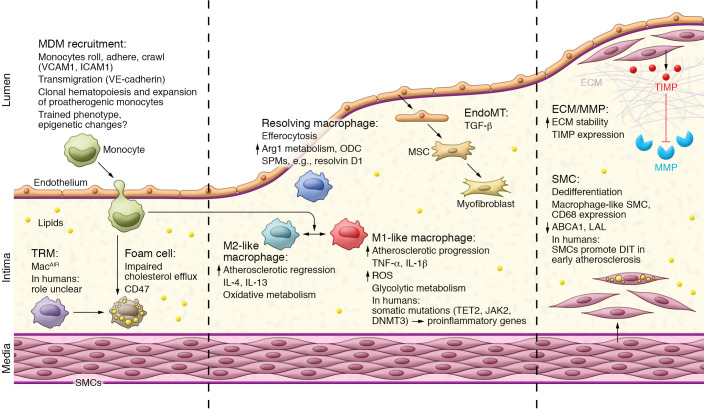
Cardiovascular disease (CVD) accounts for almost half of all deaths related to non-communicable disease worldwide, making it the single largest global cause of mortality. Although the risk factors for coronary artery disease - the most common cause of CVD - are well known and include hypertension, high cholesterol, age, and genetics, CVDs are now recognized as chronic inflammatory conditions. Arterial blockages, known as atherosclerosis, develop due to excess cholesterol accumulating within the arterial wall, creating a perpetually inflammatory state. The normally quiescent intimal layer of the vessel wall becomes laden with inflammatory cells, which alters the surrounding endothelial, smooth muscle, and extracellular matrix components to propagate disease. Macrophages, which can be either tissue resident or monocyte derived, are a key player in atherosclerotic disease progression and regression, and the understanding of their functions and origins continues to evolve with the use of deep phenotyping methodologies. This Review outlines how macrophages interact with each layer of the developing atherosclerotic plaque and discusses new concepts that are challenging our previous views on how macrophages function and our evolving understanding of the contribution of macrophages to disease.


Conflict of interest statement

Conflict of interest: The authors have declared that no conflict of interest exists.

Figures

Figure 1
Figure 1. Overview of macrophage function through the stages of atherosclerosis.
In the early stages of atherosclerosis, when cholesterol is abundant in the intima, MDMs are recruited via endothelial interactions and differentiation and, together with TRMs, engulf excess lipids to become foam cells. Certain macrophages adopt a proinflammatory M1-like phenotype that promotes inflammation and the formation of a necrotic core. During disease progression, endothelial cells can undergo EndoMT, and SMCs dedifferentiate into macrophage-like SMCs to become foam cells, all of which contribute to the growing plaque. To accommodate the growth in plaque size, ECM remodeling occurs through MMPs and, if the ECM is reduced and the SMC fibrous cap thins, plaques are prone to rupture. During disease regression and if cholesterol metabolism and efflux are efficient, macrophages take on a pro-resolving M2-like phenotype. An increase in M2-like macrophages alongside SPMs promotes inflammation resolution and plaque regression.

References

    1. Rasheed A, Rayner KJ. Macrophage responses to environmental stimuli during homeostasis and disease. Endocr Rev. 2021;42(2):407–435. - PMC - PubMed
    1. Williams NC, O’Neill LAJ. A role for the Krebs cycle intermediate citrate in metabolic reprogramming in innate immunity and inflammation. Front Immunol. 2018;9:141. doi: 10.3389/fimmu.2018.00141. - DOI - PMC - PubMed
    1. Mosser DM, Edwards JP. Exploring the full spectrum of macrophage activation. Nat Rev Immunol. 2008;8(12):958–969. doi: 10.1038/nri2448. - DOI - PMC - PubMed
    1. Joseph SB, et al. LXR-dependent gene expression is important for macrophage survival and the innate immune response. Cell. 2004;119(2):299–309. doi: 10.1016/j.cell.2004.09.032. - DOI - PubMed
    1. Takayanagi H, et al. Induction and activation of the transcription factor NFATc1 (NFAT2) integrate RANKL signaling in terminal differentiation of osteoclasts. Dev Cell. 2002;3(6):889–901. doi: 10.1016/S1534-5807(02)00369-6. - DOI - PubMed

Publication types

Grants and funding