Comment
. 2022 May 2;132(9):e159472.
doi: 10.1172/JCI159472.

Labile iron accumulation augments T follicular helper cell differentiation

Affiliations
Comment

Labile iron accumulation augments T follicular helper cell differentiation

Yogesh Scindia et al. J Clin Invest. .

Abstract

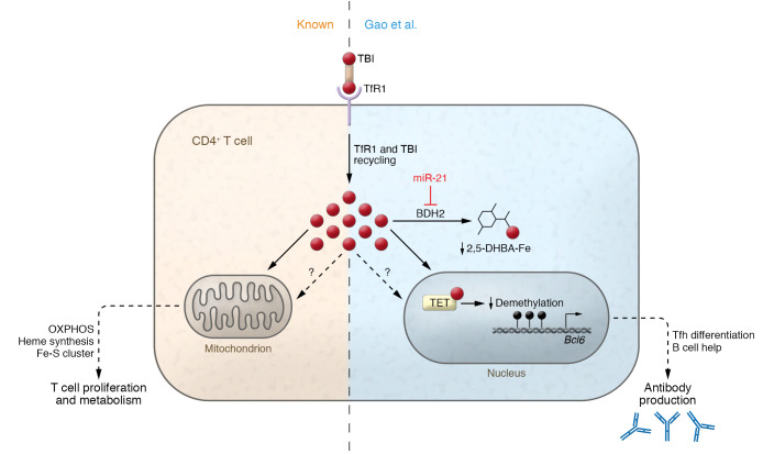
T follicular helper (Tfh) cells are a subset of CD4+ T cells that are essential in the pathogenesis of systemic lupus erythematosus (SLE). Notably, iron is required for activated CD4+ T lymphocytes to sustain high proliferation and metabolism. In this issue of the JCI, Gao et al. showed that CD4+ T cells from patients with SLE accumulated iron, augmenting their differentiation into Tfh cells and correlating with disease activity. Using human cells and murine models, the authors demonstrated that miR-21 was overexpressed in lupus T cells and inhibited 3-hydroxybutyrate dehydrogenase-2 (BDH2). The subsequent loss of BDH2 drove labile iron to accumulate in the cytoplasm and promoted TET enzyme activity, BCL6 gene demethylation, and Tfh cell differentiation. This work identifies a role for iron in CD4+ T cell biology and the development of pathogenic effectors in SLE. We await future investigations that could determine whether modulating iron levels could regulate Tfh cells in human health and disease.


Conflict of interest statement

Conflict of interest: The authors have declared that no conflict of interest exists.

Figures

Figure 1
Figure 1. The miR-21/BDH2/Fe axis promotes DNA hydroxymethylation of the BCL6 gene by regulating intracellular iron.
Activated CD4+ T cells rapidly upregulate iron import machinery, increasing transferrin receptor 1 (TfR1) to support enhanced proliferative and metabolic requirements. Gao et al. (8) identified an additional role for iron in lupus CD4+ T cell differentiation and effector function. In CD4+ T cells from lupus models, miR-21 inhibited BDH2, the hydrogenase required for the synthesis of 2,5-DHBA, a cytoplasmic iron-binding molecule that safely assimilates excess iron. Loss of 2,5-DHBA increased the availability of Fe2+ in activated CD4+ T cells. Excess iron bound to and increased the activity of TET enzyme. This led to reduced DNA demethylation and enhanced gene expression of BCL6, which encodes the Tfh cell lineage defining transcription factor. Free iron may also influence parallel pathways. Notably, increased Tfh cells worsen lupus outcomes by helping B cells make more autoantibodies. TBI, transferrin-bound iron.

Comment on

References

    1. Cronin SJF, et al. The role of iron regulation in immunometabolism and immune-related disease. Front Mol Biosci. 2019;6:116. doi: 10.3389/fmolb.2019.00116. - DOI - PMC - PubMed
    1. Ganz T, Nemeth E. Iron homeostasis in host defence and inflammation. Nat Rev Immunol. 2015;15(8):500–510. doi: 10.1038/nri3863. - DOI - PMC - PubMed
    1. Batista A, et al. Recruitment of transferrin receptor to immunological synapse in response to TCR engagement. J Immunol. 2004;172(11):6709–6714. doi: 10.4049/jimmunol.172.11.6709. - DOI - PubMed
    1. Vanoaica L, et al. Conditional deletion of ferritin h in mice reduces B and T lymphocyte populations. PLoS One. 2014;9(2):e89270. doi: 10.1371/journal.pone.0089270. - DOI - PMC - PubMed
    1. Wang Z, et al. Iron drives T helper cell pathogenicity by promoting RNA-binding protein PCBP1-mediated proinflammatory cytokine production. Immunity. 2018;49(1):80–92. doi: 10.1016/j.immuni.2018.05.008. - DOI - PubMed