Review
. 2022 Dec;16(1):25-64.
doi: 10.1080/19336918.2022.2055520.

Unravelling cell migration: defining movement from the cell surface

Affiliations
Review

Unravelling cell migration: defining movement from the cell surface

Francisco Merino-Casallo et al. Cell Adh Migr. 2022 Dec.

Abstract

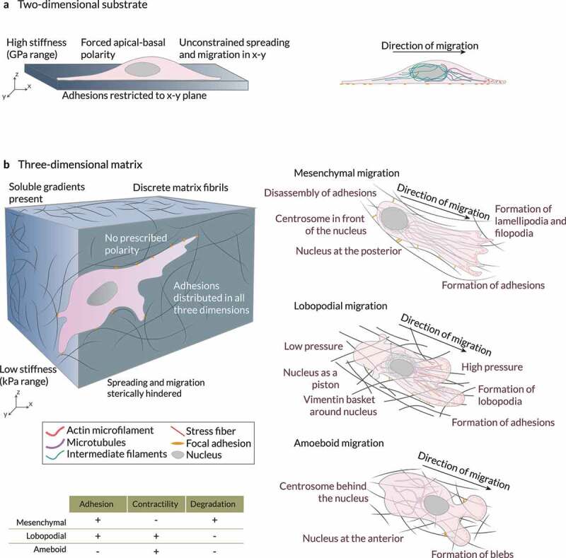
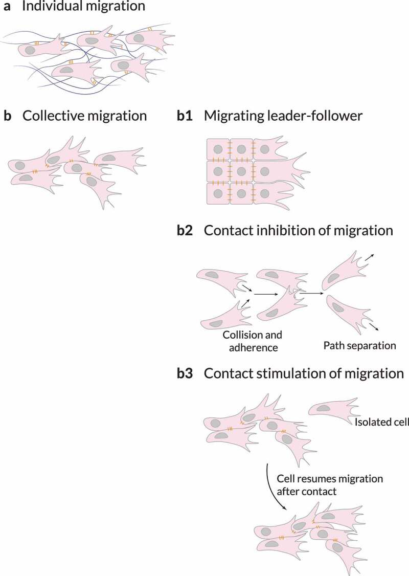
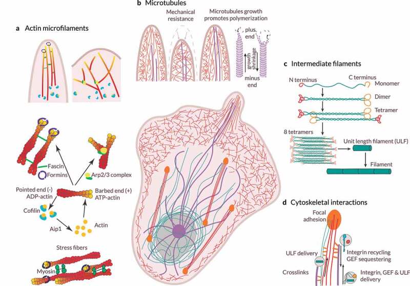
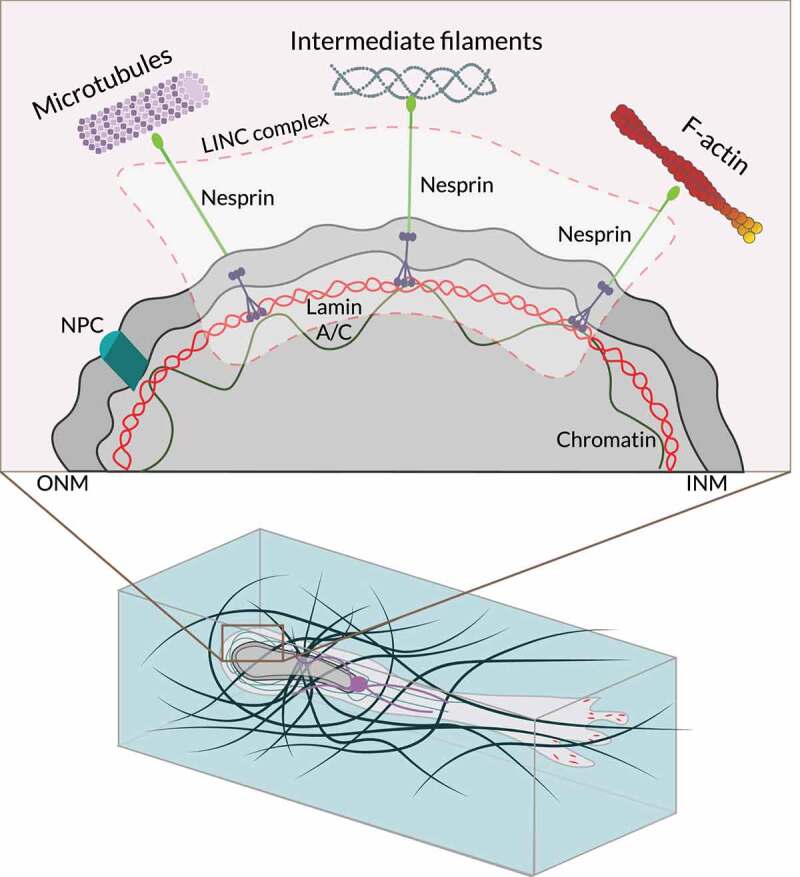
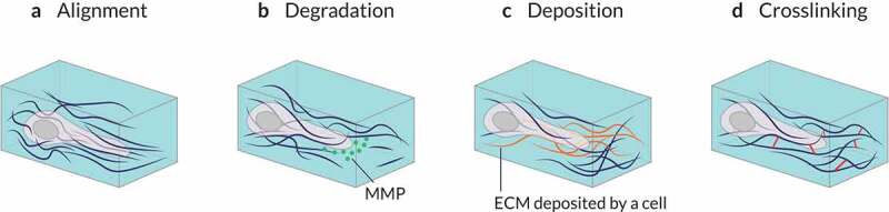
Cell motility is essential for life and development. Unfortunately, cell migration is also linked to several pathological processes, such as cancer metastasis. Cells' ability to migrate relies on many actors. Cells change their migratory strategy based on their phenotype and the properties of the surrounding microenvironment. Cell migration is, therefore, an extremely complex phenomenon. Researchers have investigated cell motility for more than a century. Recent discoveries have uncovered some of the mysteries associated with the mechanisms involved in cell migration, such as intracellular signaling and cell mechanics. These findings involve different players, including transmembrane receptors, adhesive complexes, cytoskeletal components , the nucleus, and the extracellular matrix. This review aims to give a global overview of our current understanding of cell migration.

Keywords: Cell migration; cell mechanics; extracellular matrix.


Conflict of interest statement

No potential conflict of interest was reported by the author(s).

Figures

Figure 1.
Figure 1.
Cells in 2D vs 3D Cells in (a) 2D and (b) 3D microenvironments interact differently with their surroundings. Three modes of 3D migration have been identified so far: mesenchymal, amoeboid, and lobopodial migration. In mesenchymal migration, cells attach very strongly to the extracellular matrix through mature stress fiber-linked focal adhesions. These cells also exhibit a high matrix-degrading activity. The centrosome is in front of the nucleus and the cytoskeletal networks are polarized in the direction of migration. In contrast, amoeboid migration involves very few adhesions and low protease activity. Cells migrate through the formation of contraction-based blebs or use actin-driven protrusions to glide on the substrate. The centrosome is usually behind the nucleus during amoeboid migration. Lastly, during lobopodial migration, tightly adherent cells use actomyosin contractility, hydrostatic pressure, and nuclear pistoning to form bleb-like blunt protrusions called lobopodia. These cells exhibit very low protease activity. Adapted from Refs. [39,167].
Figure 2.
Figure 2.
Sensing biochemical cues The surface abundance and distribution of transmembrane receptors, such as receptor tyrosine kinase (RTK) and G protein-coupled receptor (GPCR), is a key regulatory step. Locally high surface levels of an individual surface receptor may promote homodimerization and/or clustering, and high surface abundance of two or more of these receptors may also increase heterodimer pairing. Distinct domains within the plasma membrane, as well as the closely apposed and dynamic cortical actin cytoskeleton, affect this key step in receptor activation. The surface abundance of transmembrane receptors is predominantly controlled by receptor endocytosis, which ultimately leads to receptor degradation or recycling. When localized in specific plasma membrane domains, stimulated (ligand bound) or unstimulated (unbound) surface receptors are endocytosed or sequestered. Adapted from Ref. [66].
Figure 3.
Figure 3.
Extrinsic regulators of 3D cell migration. Different properties of the surrounding microenvironment can regulate or modulate cell migration. (a) The concentration of each extracellular matrix (ECM) component can vary locally creating, for example, gradients of stiffness (durotaxis) or ligand concentration (haptotaxis), biasing cell motility. (b) The presence and size of pores within the 3D environment – which can be altered by ECM crosslinking and may be dependent on ECM or tissue stiffness – and the level of confinement of cells mediated by the ECM modulate spatial obstruction of the substrate (steric hindrance) to cell migration. (c) Local remodeling (e.g., by proteases or local force causing physical displacement of ECM components) can also influence cell motion. The features of the local microenvironment can be overlapping; for example, increasing the concentration of ECM components can increase local stiffness and alter the sizes of pores. Adapted from Ref. [4].
Figure 4.
Figure 4.
Sensing biophysical cues by means of the integrin dynamics. On the plasma membrane, different factors (e.g., the forces from the ECM) enable integrin activation and increased affinity for ligand binding. Inside-out signals regulate displacement of intracellular integrin inhibitors and allow talin to bind to integrins, tightly controlling integrin affinity for ECM ligands. In fibroblasts, recruitment of focal adhesion kinase (FAK) to integrins has been suggested to precede talin recruitment. Integrin activation is also promoted by an outside-in mechanism through ECM binding and force application that slows the diffusion of integrin dimers within the plasma membrane. Force application leads to integrin clustering and the initiation of integrin downstream signaling through the coupling of integrins via talin and vinculin to the actin cytoskeleton. Reciprocally, actin can pull on integrins, further contributing to force generation. In fibroblasts, focal adhesions can mature further to fibrillar adhesions where talin is replaced by tensin. Trafficking of integrins regulates their availability at the plasma membrane. Integrins are constantly endocytosed from the plasma membrane. They are then efficiently recycled, with a small subset of the receptors targeted to lysosomal degradation. Integrins can be endocytosed via multiple different routes depending on the cell type, adhesion status, and cellular signaling pathways that are activated. Force regulates integrin properties. Integrin–ligand binding follows a catch bond behavior. When force (F) applied to the ligand-bound integrin is below the optimal bond force (FB), the strength (lifetime) of the bond increases with force. When F exceeds FB, the bond lifetime decays with force. Mechanical force (F) acting on integrins through their ligands can favor integrin unbending and subsequent activation, thereby triggering outside-in integrin signaling. Activation increases catch bond behavior, further strengthening the bond. If a given F is applied to an adhesion site, further integrin clustering decreases the force applied to individual integrin dimers. This minimizes elastic energy since it decreases the applied strain, and could thus be promoted. Adapted from Ref. [120].
Figure 5.
Figure 5.
Cell adhesions. Non-migratory cells must be stimulated to migrate by transcription factors, growth factors, chemokines or physical forces. (a) They can migrate as loose cohorts of individually migrating cells. (b) Cells can also maintain cohesiveness by adherence using cell–cell adhesion molecules. (b1) When migrating collectively, cells can organize into leaders and followers, in which the leaders – established by signaling cues (for example, by diffusible growth and other factors) or by mechanical cues such as those generated by neighboring cells – provide guidance as long as the biochemical or biophysical signaling is maintained. Cohesive migration of cell populations can be supported by two types of cell–cell interactions: contact inhibition of migration or contact stimulation of migration. (b2) Contact inhibition of migration is a mechanism in which colliding cells migrate in new directions after collision rather than cohering; however, on a population level, this mechanism ensures that cells maintain similar polarities, thereby resulting in directionality of migration in collectives. (b3) Contact stimulation of migration provides a direct mechanism for maintaining cohesion. In this case, cells that migrate away from contact with their neighbors cease migrating and resume migration only after being contacted by another migrating cell. Adapted from Ref. [4].
Figure 6.
Figure 6.
Cytoskeletal dynamics. (a) Assembly and organization of the actin microfilament network. The Arp2/3 complex nucleate branched actin microfilaments. Conversely, unbranched filaments may be nucleated de novo by the formins or generated from a preexisting arp2/3-nucleated network. Actin filaments grow toward the plasma membrane, generating forces that move forward the leading edge. In filopodia, fascin is the main actin microfilament cross-linking/bundling protein. Cofilin triggers actin microfilament disassembly. (b) Microtubule structure and functions. Microtubules are anchored at the centrosome and grow toward the cell cortex. Microtubule stiffness paired with the viscosity of the cytoplasm allows them to resist large compressive forces. Microtubule assembly and disassembly result in pushing and pulling forces. Stiff microtubules may provide mechanical support against membrane retraction when actin polymerization is driving membrane protrusion. Also, the growth of microtubules leads to actin polymerization in protrusions. The binding of actin microfilaments and microtubules through crosslinks allows actin microfilaments to guide microtubule growth toward focal adhesions. (c) Organization and assembly of intermediate filaments. Monomers associate to form dimers, dimers then associates to form a staggered tetramer, eight tetramers associate to form a unit-length filament (ULF), ULFs anneal to form a thick filament, and further annealing of ULFs results in filament elongation, which is followed by compaction to achieve the final intermediate filament. By organizing into a cytoplasmic nuclear cage, intermediate filaments protect the nucleus against compressive forces. Intermediate filaments also provide mechanical support for the plasma membrane in contact sites with other cells and the ECM. (d) Cytoskeletal interactions. Both actin microfilaments and microtubules can act as transport tracks of ULFs and bind to intermediate filaments through crosslinks. Further, microtubules act as transport tracks, enabling the delivery and recycling or sequestering of integrins and other signaling molecules, such as guanine nucleotide exchange factors (GEFs). As a result, microtubules regulate different processes, such as mechanotransduction and actomyosin contractility.
Figure 7.
Figure 7.
The nucleus during 3D cell migration. Mesenchymal cell migration within the extracellular matrix (ECM) requires multiple steps, including nuclear rotation and repositioning. Translocating the bulky nucleus of migrating cells through ECM barriers can become challenging unless the ECM is loose or highly pliable. Alternatively, the nucleus can be used to drive lobopodial cell migration, acting as a pressure-generating piston. Also, during amoeboid migration, cells can use the nucleus as a mechanical gauge or ruler by presenting it anteriorly to ‘measure’ the diameter of pores or passages in the ECM microenvironment. The cell then translocates through a passageway that is sufficiently wide to accommodate the bulky nucleus as the route of least resistance. The LINC complex is at the center of the nuclear-cytoskeletal coupling. On the cytoplasmic side, different nesprin isoforms connect the nucleus to the cytoskeleton. In the perinuclear space, nesprins bind SUN proteins, which span the inner nuclear membrane (INM) and interact with the nuclear lamina through lamin A. Emerin, a protein from the inner nuclear membrane, anchors SUN protein to lamin A and interacts directly with chromatin. NPC, nuclear pore complex. ONM, outer nuclear membrane. Adapted from Ref. [133].
Figure 8.
Figure 8.
Matrix remodeling through cell–matrix interactions. (a) Mechanical forces exerted by cells can structurally remodel the surrounding matrix by stretching and aligning fibers of the extracellular matrix (ECM). (b) Cells may biochemically degrade a surrounding ECM by secreting various types of matrix metalloproteinases (MMPs). (c) Specific types of cells, such as fibroblasts, deposit additional ECM components on the surrounding matrix. This can lead to elevated matrix stiffness and smaller matrix pore size. (d) Cells can cross-link matrix fibers, resulting in the enhanced stiffness and elasticity of the ECM.

References

    1. Karamanos NK, Theocharis AD, Piperigkou Z, et al. A guide to the composition and functions of the extracellular matrix. FEBS J. 2021;228:15776. - PubMed
    1. David ACW, Kenneth MY.. The extracellular matrix in development. Development. 147(10):2020. - PMC - PubMed
    1. Marsha CL, Cynthia AR-K.. Targeting extracellular matrix stiffness to attenuate disease: from molecular mechanisms to clinical trials. Sci Transl Med. 10(422):2018. - PubMed
    1. Kenneth MY, Sixt M. Mechanisms of 3D cell migration. Nat Rev Mol Cell Biol. 2019;20(12):738–752. - PubMed
    1. Eddy CZ, Raposo H, Wong R, et al. Morphodynamics facilitate cancer cells to navigate 3D extracellular matrix. Scientific Reports. 2020;11(1):1–10. 10.1038/s41598-021-99902-9. - DOI - PMC - PubMed

Publication types

LinkOut - more resources