Review
. 2022 Jun 7;23(6):e54217.
doi: 10.15252/embr.202154217. Epub 2022 May 2.

Mechanisms of DNA damage-mediated neurotoxicity in neurodegenerative disease

Affiliations
Review

Mechanisms of DNA damage-mediated neurotoxicity in neurodegenerative disease

Gwyneth Welch et al. EMBO Rep. .

Abstract

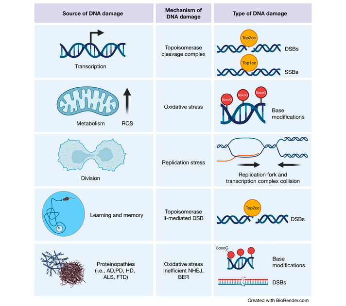
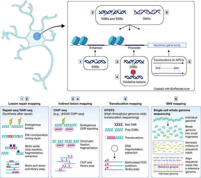
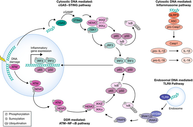
Neurons are highly susceptible to DNA damage accumulation due to their large energy requirements, elevated transcriptional activity, and long lifespan. While newer research has shown that DNA breaks and mutations may facilitate neuron diversity during development and neuronal function throughout life, a wealth of evidence indicates deficient DNA damage repair underlies many neurological disorders, especially age-associated neurodegenerative diseases. Recently, efforts to clarify the molecular link between DNA damage and neurodegeneration have improved our understanding of how the genomic location of DNA damage and defunct repair proteins impact neuron health. Additionally, work establishing a role for senescence in the aging and diseased brain reveals DNA damage may play a central role in neuroinflammation associated with neurodegenerative disease.

Keywords: DNA damage; DNA damage repair; inflammation; neurodegeneration; neuron.


Figures

Figure 1
Figure 1. Sources of DNA damage in the brain
Transcriptional activities can result in topoisomerase cleavage complexes, which lead to the induction of SSBs or DSBs depending upon the topoisomerase in question. Additionally, metabolic activity by mitochondria generate ROS, which can scar DNA bases with oxidative modifications. Although less common in the adult brain, cell division is also a source of DNA damage. Proliferation increases the chance of replication fork and transcription complex collision, thereby inducing DSBs. In the developing brain, this is a particular risk for NPCs, which harbor increased translocations in long genes (where these collisions are most likely to occur) important for neuronal function. Cognitively demanding tasks recruit specific neuronal ensembles whose plasticity is highly dependent upon immediate early gene transcription. Therefore, neurons generate topoisomerase II‐mediated DSBs in response to learning and memory. Finally, the proteins responsible for various neurodegenerative diseases have also been found to play roles in DNA damage detection and repair. (Created with BioRender.com).
Figure 2
Figure 2. DNA lesions and mutations identified in neural genes and the techniques used to map them
While all cell types incur DNA damage and mutations, neurons in particular are susceptible due to activity‐induced transcription. Immediate‐early genes and other neuronal genes that enable synaptic function are highly transcribed. Accordingly, they accumulate DNA lesions and mutations in their gene body (Wei et al, 2016) and regulatory regions (Lodato et al, ; Reid et al, ; Wu et al, 2021). The induction of DSBs in the promoters of immediate‐early genes facilitates gene expression (Madabhushi et al, 2015). Over time, these insults may impair neural function (Lu et al, ; Lodato et al, ; Pao et al, 2020). (Created with BioRender.com).
Figure 3
Figure 3. DNA damage initiates inflammatory signaling through DDR and cGAS‐STING. ATM‐NF‐kB pathway
The activation of ATM by DSBs leads to its coupling with NEMO in the nucleus. NEMO is phosphorylated by ATM and SUMOylated by PIASy in a PARP1‐dependent manner, which is not shown here. These modifications lead to NEMO monoubiquitination, and the ATM‐NEMO complex is transported to the cytoplasm. Here, NEMO partners with IKKb and IKKa to form the active Inhibitor of KappaB Kinase (IKK) complex. The IKK phosphorylates IkB, allowing NF‐kB to be transported to the nucleus. The most common form of NF‐kB is the heterodimer p50‐p65, shown here. The phosphorylation of IkB leads to its polyubiquitination and subsequent degradation. cGAS‐STING pathway: DSBs result in the leakage of self‐DNA into the cytosol, which is sensed by cGAS. cGAS generates second messenger cyclic GAMP. cGAMP binds to STING, which activates TANK‐binding kinase 1 (TBK1), which in turn activates IFN Regulatory Factor 3 (IRF3). Homodimerized IRF3 transports to the nucleus and activates the expression of inflammatory genes. STING also facilitates the formation of the IKK complex, which phosphorylates IkB to activate NF‐kB. TLR9 pathway: Endosomal double‐strand DNA is bound by TLR9, activating MyD88, which interacts with and activates IRAK1,2,and 4. IRAK1 and 4 dissociate from MyD88 and activate TRAF6. TRAF6 ubiquitinates NEMO, a member of the IKK complex that results in NF‐kB translocation into the nucleus. Inflammasome pathway: NLRP3 detects cytosolic DNA, leading to the assembly of the NLRP3, ASC, pro‐Caspase I inflammasome. pro‐Caspase I autoproteolytically matures to functional Caspase I, which cleaves pro‐IL1b and pro‐IL18 to generate functional IL1b and IL18.

References

    1. Abe T, Barber GN (2014) Cytosolic‐DNA‐mediated, STING‐dependent proinflammatory gene induction necessitates canonical NF‐κB activation through TBK1. J Virol 88: 5328–5341 - PMC - PubMed
    1. Adamec E, Vonsattel JP, Nixon RA (1999) DNA strand breaks in Alzheimer’s disease. Brain Res 849: 67–77 - PubMed
    1. Adamowicz M, Hailstone R, Demin AA, Komulainen E, Hanzlikova H, Brazina J, Gautam A, Wells SE, Caldecott KW (2021) XRCC1 protects transcription from toxic PARP1 activity during DNA base excision repair. Nat Cell Biol 23: 1287–1298 - PMC - PubMed
    1. Aditi , Downing SM, Schreiner PA, Kwak YD, Li Y, Shaw TI, Russell HR, McKinnon PJ (2021) Genome instability independent of type I interferon signaling drives neuropathology caused by impaired ribonucleotide excision repair. Neuron 109: 3962–3979.e6 - PMC - PubMed
    1. Ahn J, Gutman D, Saijo S, Barber GN (2012) STING manifests self DNA‐dependent inflammatory disease. Proc Natl Acad Sci USA 109: 19386–19391 - PMC - PubMed