. 2022 Jul 1;71(7):1579-1590.
doi: 10.2337/db21-0926.

Indirect and Direct Effects of SARS-CoV-2 on Human Pancreatic Islets

Affiliations

Indirect and Direct Effects of SARS-CoV-2 on Human Pancreatic Islets

Moufida Ben Nasr et al. Diabetes. .

Abstract

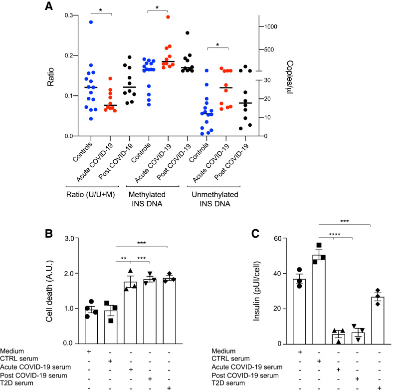
Recent studies have shown that severe acute respiratory syndrome coronavirus 2 (SARS-CoV-2) infection may induce metabolic distress, leading to hyperglycemia in patients affected by coronavirus disease 19 (COVID-19). We investigated the potential indirect and direct effects of SARS-CoV-2 on human pancreatic islets in 10 patients who became hyperglycemic after COVID-19. Although there was no evidence of peripheral anti-islet autoimmunity, the serum of these patients displayed toxicity on human pancreatic islets, which could be abrogated by the use of anti-interleukin-1β (IL-1β), anti-IL-6, and anti-tumor necrosis factor α, cytokines known to be highly upregulated during COVID-19. Interestingly, the receptors of those aforementioned cytokines were highly expressed on human pancreatic islets. An increase in peripheral unmethylated INS DNA, a marker of cell death, was evident in several patients with COVID-19. Pathology of the pancreas from deceased hyperglycemic patients who had COVID-19 revealed mild lymphocytic infiltration of pancreatic islets and pancreatic lymph nodes. Moreover, SARS-CoV-2-specific viral RNA, along with the presence of several immature insulin granules or proinsulin, was detected in postmortem pancreatic tissues, suggestive of β-cell-altered proinsulin processing, as well as β-cell degeneration and hyperstimulation. These data demonstrate that SARS-CoV-2 may negatively affect human pancreatic islet function and survival by creating inflammatory conditions, possibly with a direct tropism, which may in turn lead to metabolic abnormalities observed in patients with COVID-19.

Trial registration: ClinicalTrials.gov NCT04463849.


Figures

Figure 1
Figure 1
In vivo and in vitro evidence of cell death associated with COVID-19. A: Comparison of the ratio of unmethylated (U) INS DNA/U plus methylated (M) INS DNA was performed in patients with COVID-19 (acute COVID-19), in patients who had recovered from COVID-19 (post COVID-19), and in healthy control subjects. Ratio of 0.196 was considered as a cutoff (mean plus 2SD; 97.7th percentile) for the U/M+U calculation, with a ratio ≥0.196 indicating β-cell death. Comparison of the levels of U INS DNA as well as of M INS DNA (i.e., copies of U/μL and copies of M INS DNA/μL) was performed and measured by droplet digital PCR in serum samples from patients with COVID-19 (acute COVID-19 and post COVID-19) and in healthy control subjects. B and C: Rates of cell death and insulin secretion were analyzed for purified human islets upon in vitro challenge with serum from healthy control subjects, from patients with COVID-19 (acute COVID-19), from patients who had recovered from COVID-19 (post COVID-19), or from patients with type 2 diabetes (T2D). Data are representative of n = 15 samples from the subgroup of healthy control subjects and n = 10 from acute COVID-19 and from post COVID-19. Data are representative of a pool of n = 3–4 commercial islet preparations in three separate experiments (n = 3–5 pooled sera tested). Data are represented as mean ± SEM. Ordinary one-way ANOVA test with Bonferroni correction was used for calculating statistical significance between all groups. *P < 0.05, **P < 0.01, ***P < 0.001, ****P < 0.0001. AU, arbitrary unit.
Figure 2
Figure 2
Altered COVID-19–associated secretome has a toxic effect on human pancreatic islets. A: Receptome analysis of human pancreatic islets depicting the expression of proinflammatory cytokine receptors (TNAFAR, IL-13R, IL-1R1, and IL-6R); mRNAs of GLP1R, Glut-2, MAFA, GCK, and PDX-1 are shown as well. Data are representative of four independent human islet isolations (n = 4 donors), represented as mean plus SD. B and C: Rates of cell death (B) and insulin secretion (C) were analyzed for purified human pancreatic islets upon in vitro challenge with a selected panel of cytokines (IL-1β, IL-6, IL-13, IP-10, and TNF-α) observed to be upregulated in the serum of patients with COVID-19. Effects were assessed when the cytokines were added alone or in combination. D and E: Rates of cell death (D) and insulin secretion (E) were analyzed for purified human pancreatic islets upon in vitro challenge with serum isolated from a patient who recovered from COVID-19 (post COVID-19) in the presence of neutralizing antibodies (anti–IL-1β, anti–IL-6, or anti–IL-13) alone or in combination, as compared with when challenged with serum from healthy control subjects. Data are representative of a pool of n = 3–4 commercial islet preparations in three separate experiments (n = 3–5 pooled sera tested). Data are represented as mean ± SEM. Ordinary one-way ANOVA test with Bonferroni correction was used for calculating statistical significance between all groups. *P < 0.05, **P < 0.01, ***P < 0.001, ****P < 0.0001. AU, arbitrary unit; RPKM, reads per kilobase per million mapped reads.
Figure 3
Figure 3
Abnormalities in the endocrine pancreas are observed in patients with COVID-19. A: Histologic examination of pancreatic section retrieved from a patient with COVID-19 stained with H-E showing mild islet lymphocytic infiltration. B: Bar graph showing the results of a SARS-CoV-2 RT-PCR assay using the 2019-nCoV_N1 and 2019-nCoV_N2 primer probe sets performed on RNA samples extracted from pancreatic sections from patients with COVID-19 (P1, P2, P3, and P4) and from healthy control subjects, showing detectable viral RNA in patient P1. RNA extracted from a Centers for Disease Control and Prevention (CDC)–positive control DNA plasmid (CDC+) was used as a positive control, and RNA extracted from a CDC-negative sample (CDC) was used as a negative control. Cycle threshold (CT) values are shown. C: H-E staining of PLNs from a patient with COVID-19. D: Bar graph depicting the results of a SARS-CoV-2 RT-PCR assay using the 2019-nCoV_N1 and 2019-nCoV_N2 primer probe sets performed on RNA samples extracted from PLNs from a patient with COVID-19 (P1), showing detectable viral RNA in patient P1. RNA extracted from a CDC+ control DNA plasmid was used as a positive control, and RNA extracted from a CDC sample was used as a negative control. CT values are shown. Magnification in panels A and D, ×10 and panel C, ×20; scale bars in panels A and D, 100 μm and panel C, 100 μm. EJ: Transmission electron microscopic analysis of pancreatic tissue from patients with COVID-19 as compared with healthy control subjects and patients with type 2 diabetes, showing similar islet alterations in pancreatic tissues from patients with COVID-19 and patients with type 2 diabetes; arrows indicate the presence of insulin secretion granules; in healthy control subjects, arrows delineate insulin granules with different degrees of granulation; in diabetic samples as well as in COVID-19 samples, arrows delineate β-cells with several immature granules. Asterisks in panels H, I, and J show mature insulin granules. K: Quantification of the proportions of immature insulin granules per total mature insulin secretion granules; n = 3 cases per section were analyzed. L and M: Transmission electron microscopic analysis of pancreatic tissue from a patient with COVID-19 as compared with that from healthy control subjects depicting the presence of several vacuoles (shown by black arrows) in the vicinity of β-cells from a patient with COVID-19. N: SARS-CoV-2 spike S1 staining depicted within endocrine cells; arrows indicate the positive staining within endocrine cells of the pancreas; asterisk refers to an islet, and arrowheads indicate pancreatic acinar cells. AU, arbitrary unit.
Figure 4
Figure 4
SARS-CoV-2 spike protein S1 localizes within endocrine pancreatic β-cells and exocrine pancreatic cells. AI: Confocal microscopic analysis of pancreatic tissue from a patient with COVID-19 depicting the localization of SARS-CoV-2 spike protein S1 within β-cells (insulin-positive cells) (shown in panel C) and within exocrine cells, trypsin-positive cells (shown in panel F), and CK19-positive cells (as shown in I). Scale bars in panels AI, 10 μm.

References

    1. Starling S. How COVID-19 disrupts glycometabolic control. Nat Rev Endocrinol 2021;17:448 - PMC - PubMed
    1. Montefusco L, Ben Nasr M, D’Addio F, et al. . Acute and long-term disruption of glycometabolic control after SARS-CoV-2 infection. Nat Metab 2021;3:774–785 - PMC - PubMed
    1. Wu CT, Lidsky PV, Xiao Y, et al. . SARS-CoV-2 infects human pancreatic β cells and elicits β cell impairment. Cell Metab 2021;33:1565–1576.e5 - PMC - PubMed
    1. Solerte SB, D’Addio F, Trevisan R, et al. . Sitagliptin treatment at the time of hospitalization was associated with reduced mortality in patients with type 2 diabetes and COVID-19: a multicenter, case-control, retrospective, observational study. Diabetes Care 2020;43:2999–3006 - PMC - PubMed
    1. Yang JK, Feng Y, Yuan MY, et al. . Plasma glucose levels and diabetes are independent predictors for mortality and morbidity in patients with SARS. Diabet Med 2006;23:623–628 - PubMed

Publication types