

A Review of Treatment for Acute and Chronic Pars Fractures in the Lumbar Spine
- PMID: 35499747
- PMCID: PMC9276862
- DOI: 10.1007/s12178-022-09760-9
A Review of Treatment for Acute and Chronic Pars Fractures in the Lumbar Spine
Abstract
Purpose of review: Spondylolysis remains one of the most common causes of lower back pain in the pediatric and adolescent populations and is particularly prevalent in young sporting individuals. Despite this, approaches to diagnostic imaging and both conservative and surgical treatment vary widely among surgeons. The current review investigates recent literature on the etiology, clinical presentation, diagnosis, and treatment of spondylolysis. In particular, it interrogates the use of various advanced imaging modalities (CT, MRI, SPECT) in diagnosis as well as common surgical approaches to the condition.
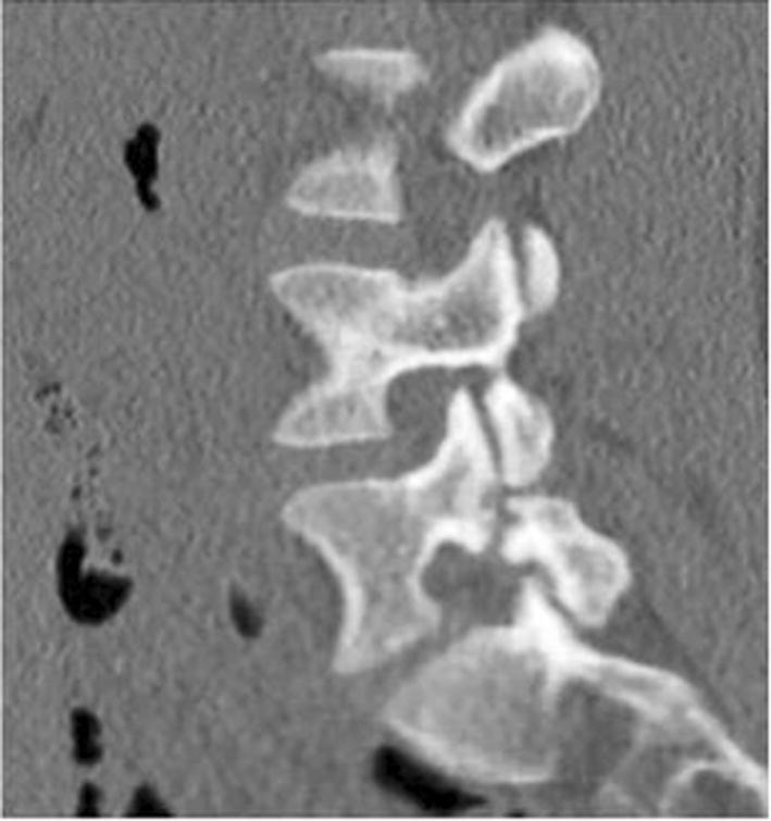
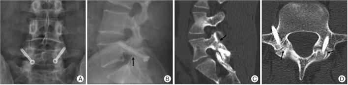
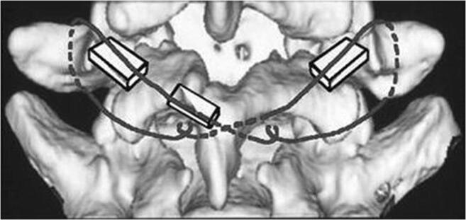
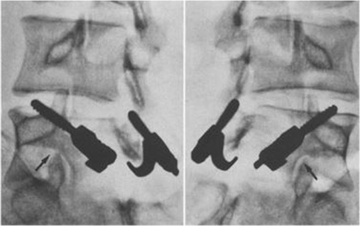
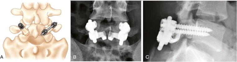
Recent findings: Recent data has provided more information on how pars defect laterality, stage, and presence or absence of bone marrow edema impact healing potential. Other studies have highlighted the advantages of using MRI for spondylolysis diagnosis. Other data has provided more clarity on which adults may benefit from direct pars repair, while other studies have compared the various techniques for direct repair of pars defects. While the exact cause of spondylolysis remains unclear, there is growing understanding of the behavioral, genetic, and biomechanical risk factors that predispose individuals to the condition. MRI may be emerging as the advanced imaging modality of choice for diagnosis due to its lack of radiation and comparable sensitivity to other advanced imaging techniques. Conservative treatment remains the first step in management due to excellent outcomes in most patients, with surgical intervention rarely necessary. In patients that do require surgery, direct repair using a pedicle screw-based approach is preferred over spinal fusion and other direct repair techniques.
Keywords: Athletics; Pars Defect; Spine surgery; Spondylolisthesis; Spondylolysis.
© 2022. The Author(s), under exclusive licence to Springer Science+Business Media, LLC, part of Springer Nature.
Conflict of interest statement
Alexander Linton—No conflicts. Dr. Wellington Hsu—Advisory board member of Stryker, Medtronic, Asahi, Bioventus.
Figures
References
-
- Hu SS, Tribus CB, Diab M, Ghanayem AJ. Spondylolisthesis and spondylolysis. Instr Course Lect. 2008;57:431–445. - PubMed
Publication types
LinkOut - more resources
Full Text Sources
Research Materials
Miscellaneous
