

Specific Detection of Acanthamoeba species using Polyclonal Peptide Antibody Targeting the Periplasmic Binding Protein of A. castellanii
- PMID: 35500897
- PMCID: PMC9058276
- DOI: 10.3347/kjp.2022.60.2.143
Specific Detection of Acanthamoeba species using Polyclonal Peptide Antibody Targeting the Periplasmic Binding Protein of A. castellanii
Abstract
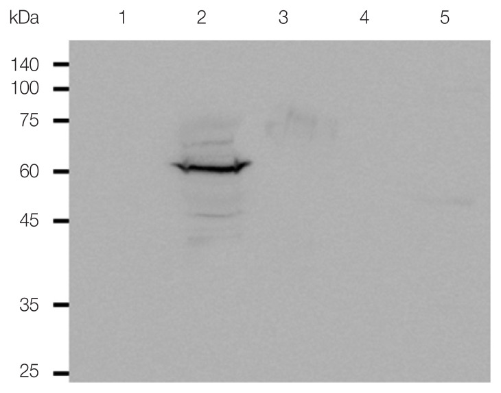
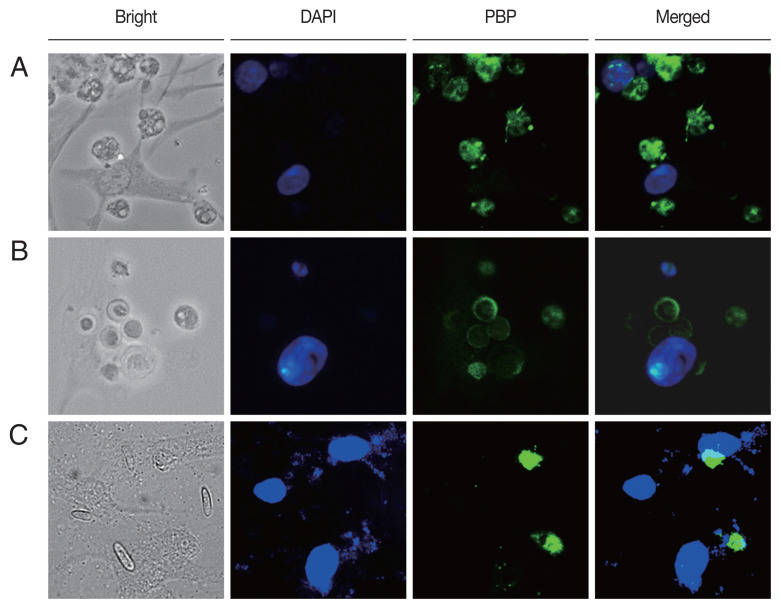
Acanthamoeba keratitis (AK) is a rare ocular disease, but it is a painful and sight-threatening infectious disease. Early diagnosis and adequate treatment are necessary to prevent serious complications. While AK is frequently diagnosis via several PCR assays or Acanthamoeba-specific antibodies, a more specific and effective diagnostic method is required. This study described the production of a polyclonal peptide antibody against the periplasmic binding protein (PBP) of A. castellanii and investigated its diagnostic potential. Western blot analysis showed that the PBP antibody specifically reacted with the cell lysates of A. castellanii. However, the PBP antibody did not interact with human corneal epithelial (HCE) cells and the other 3 major causative agents of keratitis. Immunocytochemistry (ICC) results revealed the specific detection of A. castellanii trophozoites and cysts by PBP antibodies when A. castellanii were co-cultured with HCE cells. PBP antibody specificity was further confirmed by co-culture of A. castellanii trophozoites with F. solani, S. aureus, and P. aeruginosa via ICC. The PBP antibody specifically reacted with the trophozoites and cysts of A. polyphaga, A. hatchetti, A. culbertsoni, A. royreba, and A. healyi, thus demonstrated its genus-specific nature. These results showed that the PBP polyclonal peptide antibody of A. castellanii could specifically detect several species of Acanthamoeba, contributing to the development of an effective antibody-based AK diagnostics.
Keywords: Acanthamoeba keratitis; peptide antibody; periplasmic binding protein; species specificity.
Conflict of interest statement
The authors declare no conflict of interest related to this study.
Figures

Similar articles
-
Evaluating the Diagnostic Potential of Chorismate Mutase Poly-Clonal Peptide Antibody for the Acanthamoeba Keratitis in an Animal Model.Pathogens. 2023 Mar 28;12(4):526. doi: 10.3390/pathogens12040526. Pathogens. 2023. PMID: 37111412 Free PMC article.
-
Characterization of a Peptide Antibody Specific to the Adenylyl Cyclase-Associated Protein of Acanthamoeba castellanii.Korean J Parasitol. 2022 Feb;60(1):7-14. doi: 10.3347/kjp.2022.60.1.7. Epub 2022 Feb 23. Korean J Parasitol. 2022. PMID: 35247949 Free PMC article.
-
Detection of Acanthamoeba spp. using carboxylesterase antibody and its usage for diagnosing Acanthamoeba-keratitis.PLoS One. 2022 Jan 5;17(1):e0262223. doi: 10.1371/journal.pone.0262223. eCollection 2022. PLoS One. 2022. PMID: 34986189 Free PMC article.
-
Chorismate mutase peptide antibody enables specific detection of Acanthamoeba.PLoS One. 2021 Apr 23;16(4):e0250342. doi: 10.1371/journal.pone.0250342. eCollection 2021. PLoS One. 2021. PMID: 33891646 Free PMC article.
-
The biology of Acanthamoeba keratitis.Exp Eye Res. 2021 Jan;202:108365. doi: 10.1016/j.exer.2020.108365. Epub 2020 Nov 19. Exp Eye Res. 2021. PMID: 33221372 Free PMC article. Review.
Cited by
-
Evaluating the Diagnostic Potential of Chorismate Mutase Poly-Clonal Peptide Antibody for the Acanthamoeba Keratitis in an Animal Model.Pathogens. 2023 Mar 28;12(4):526. doi: 10.3390/pathogens12040526. Pathogens. 2023. PMID: 37111412 Free PMC article.
-
Evaluation of the potential for diagnosis of fungal keratitis using a Fusarium-specific antibody.Sci Rep. 2025 Jul 1;15(1):21583. doi: 10.1038/s41598-025-08719-3. Sci Rep. 2025. PMID: 40595346 Free PMC article.
-
Detection of Acanthamoeba from Acanthamoeba Keratitis Mouse Model Using Acanthamoeba-Specific Antibodies.Microorganisms. 2022 Aug 25;10(9):1711. doi: 10.3390/microorganisms10091711. Microorganisms. 2022. PMID: 36144313 Free PMC article.
-
Detection of Fusarium solani using cutinase antibody and its application in diagnosing fungal keratitis in an animal model.PLoS One. 2025 Aug 20;20(8):e0330455. doi: 10.1371/journal.pone.0330455. eCollection 2025. PLoS One. 2025. PMID: 40834005 Free PMC article.
References
MeSH terms
Substances
Grants and funding
LinkOut - more resources
Full Text Sources
Research Materials
Miscellaneous
