Review
. 2022 Sep;14(5):e1807.
doi: 10.1002/wnan.1807. Epub 2022 May 2.

Multimodality imaging of nanoparticle-based vaccines: Shedding light on immunology

Affiliations
Review

Multimodality imaging of nanoparticle-based vaccines: Shedding light on immunology

Muhsin H Younis et al. Wiley Interdiscip Rev Nanomed Nanobiotechnol. 2022 Sep.

Abstract

In recent years, there have been significant innovations in the development of nanoparticle-based vaccines and vaccine delivery systems. For the purposes of both design and evaluation, these nanovaccines are imaged using the wealth of understanding established around medical imaging of nanomaterials. An important insight to the advancement of the field of nanovaccines can be given by an analysis of the design rationale of an imaging platform, as well as the significance of the information provided by imaging. Nanovaccine imaging strategies can be categorized by the imaging modality leveraged, but it is also worth understanding the superiority or convenience of a given modality over others in a given context of a particular nanovaccine. The most important imaging modalities in this endeavor are optical imaging including near-infrared fluorescence imaging (NIRF), emission tomography methods such as positron emission tomography (PET) and single photon emission computed tomography (SPECT) with or without computed tomography (CT) or magnetic resonance (MR), the emerging magnetic particle imaging (MPI), and finally, multimodal applications of imaging which include molecular imaging with magnetic resonance imaging (MRI) and photoacoustic (PA) imaging. One finds that each of these modalities has strengths and weaknesses, but optical and PET imaging tend, in this context, to be currently the most accessible, convenient, and informative modalities. Nevertheless, an important principle is that there is not a one-size-fits-all solution, and that the specific nanovaccine in question must be compatible with a particular imaging modality. This article is categorized under: Nanotechnology Approaches to Biology > Nanoscale Systems in Biology Therapeutic Approaches and Drug Discovery > Nanomedicine for Oncologic Disease Therapeutic Approaches and Drug Discovery > Nanomedicine for Infectious Disease.

Keywords: fluorescence imaging; medical imaging; nanomaterials; positron emission tomography; vaccines.


Conflict of interest statement

CONFLICT OF INTEREST

Weibo Cai is a scientific advisor, stockholder, and grantee of Focus-X Therapeutics, Inc. All other authors declare that they have no conflict of interest.

Figures

FIGURE 1
FIGURE 1
The process of tumor antigen presentation which is caused by cancer vaccination. Injection of an antigen causes uptake by antigen presenting cells (APCs). APCs then travel to draining lymph nodes, wherein mature dendritic cells (DCs) present antigen-derived peptides on MHC class I molecules to CD8+ and those on MHC class II molecules to CD4+ T cells. Several factors are involved in the activation of these T cells (CD28, CD27, CD40, IL-12, and Type-I interferon). With activated, antigen-specific T cells now formed, the cancer cells can now be recognized and killed by the production of effector cytokines such as IFN-γ and tumor necrosis factor (TNF). Adapted with permission from Persano et al. (2021).
FIGURE 2
FIGURE 2
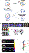
(a) A SARS-CoV-2 vaccine nanoplatform named ARCoV, wherein lipid nanoparticles are designed to carry an mRNA sequence corresponding to the receptor binding domain (RBD) of SARS-CoV-2. Immunization confers protection from SARS-CoV-2 in mice. (b) Fluorescence imaging of FLuc indicating presence of mRNA-LNP in mice, intramuscular (i.m.) and subcutaneous (s.c.) injections as well as intranasal (i.n.) inoculation. (c) Ex vivo imaging of organs from mice in the i.m. injection group (left) and free injection of LNP group (right). Adapted with permission from Zhang et al. (2020). (d) NIRF imaging of mice injected with OVA-ZW, ONP1, and ONP2, at 1 and 6 h. (e) Concentration of conjugated molecules versus time for each group. Both (d) and (e) demonstrated that OVA-ZW had higher uptake in the popliteal lymph nodes for the given time frames. Adapted with permission from Katagiri et al. (2019).
FIGURE 3
FIGURE 3
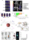
(a) PET imaging of different 89Zr-OVA-loaded formulations in mice. (b) PET/CT image showing the injection site with respect to various ROIs as well as uptake values. Adapted with permission from Han et al. (2021). (c) PET images at 6 h p.i. of CpG, PEG-CpG, MH2C, and MH3C formulations of AlbiVax. Axillary (Ax) and inguinal (In) lymph nodes are indicated in the MH3C image. (d) %ID of all formulations of AlbiVax. Adapted with permission from Zhu et al. (2017). (e) Design of PEGylated magnetite nanoparticles carrying immunogen OVA, adjuvant CpG, as well as gamma emitting radioisotope 67Ga and fluorophore DPPE-Rho. Injection in mice shows that the nanovaccine targets T-cells in lymph nodes to elicit immunogenic effect. (f) 67Ga-SPECT/CT of mice 3 h p.i. with nanovaccine formulation, injection in the forearm, (g) injection in the hock, and (h) injection in the flank. (i) Image of organs harvested from a mouse in the flank injection group 24 h p.i. (j) 67Ga-SPECT/CT of harvested organs. (k) %ID/g in various ROIs. Adapted with permission from Ruiz de-Angulo et al. (2016).
FIGURE 4
FIGURE 4
(a) A side-by-side comparison of standard MPI and “dual-color” MPI (with CT for the anatomical background), using both a lung-targeting tracer (green) and a liver-targeting tracer (red). Adapted with permission from Zhou et al. (2018). (b) MPI of mice injected with ferucarbotran-labeled tumor-effective T cells for brain tumor therapy, 24 h p.i. with intravenous (IV) injection, and intracerebroventricular (ICV) injection. Adapted with permission from Rivera-Rodriguez et al. (2021).
FIGURE 5
FIGURE 5
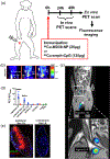
(a) Schematic of the study design wherein a nanovaccine was developed for PET/MR imaging and subsequent fluorescence imaging of a rhesus macaque model. (b) PET-only images of three rhesus macaques 24 h p.i. of 64Cu-MD39-NPs. (c) Combined PET/MR composite of two rhesus macaques 24 h p.i. 64Cu-MD39-NPs, injection site and lymph nodes are isolated in the PET scan while MRI provides anatomical context. (d) SUVmean of all three rhesus macaques, indicating high uptake in the targeted lymph nodes (LNs). (e) Ex vivo (24 h p.i.) fluorescence imaging of ipsilateral and contralateral iliac LNs of rhesus macaques injected with fluorescent MD39-NP (green), Amph-CpG (red), and CD35 (blue). Adapted with permission from Martin et al. (2021).
FIGURE 6
FIGURE 6
(a) Schematic of the study design by Zhao et al. OVA@Mn-DAP nanoparticles are formed, carrying the model antigen OVA and act as natural MRI contrast agents due to Mn2+ incorporation or fluorophores if OVA-Cy5.5 is used. Following synthesis, this nanovaccine is tested in vivo to analyze antitumor immunity. (b) Side-by-side comparison of NIRF and MRI of OVA@Mn-DAP nanovaccine in mice, time points p.i. in the footpad are above the images. Both imaging modalities demonstrate accumulation of nanovaccine in popliteal lymph nodes. Adapted from Zhao et al. (2019).
FIGURE 7
FIGURE 7
Schematic of the study by Liang et al., in which a fluorescently labeled gold nanocage-based nanovaccine (capable of both fluorescence and PA imaging) is injected in mice to deliver a melanoma antigen and adjuvant. Adapted with permission from Liang et al. (2017).

Similar articles

Cited by

References

    1. Anderson CJ, & Ferdani R (2009). Copper-64 radiopharmaceuticals for PET imaging of cancer: Advances in preclinical and clinical research. Cancer Biotherapy & Radiopharmaceuticals, 24(4), 379–393. 10.1089/cbr.2009.0674 - DOI - PMC - PubMed
    1. Bayne-Jones S (1917). Equilibria in precipitin reactions: The coexistence of a single free antigen and its antibody in the same serum. The Journal of Experimental Medicine, 25(6), 837–853. 10.1084/jem.25.6.837 - DOI - PMC - PubMed
    1. Beard P (2011). Biomedical photoacoustic imaging. Interface Focus, 1(4), 602–631. 10.1098/rsfs.2011.0028 - DOI - PMC - PubMed
    1. Beatty GL, & Gladney WL (2015). Immune escape mechanisms as a guide for cancer immunotherapy. Clinical Cancer Research, 21(4), 687–692. 10.1158/1078-0432.CCR-14-1860 - DOI - PMC - PubMed
    1. Binnewies M, Roberts EW, Kersten K, Chan V, Fearon DF, Merad M, Coussens LM, Gabrilovich DI, Ostrand-Rosenberg S, Hedrick CC, Vonderheide RH, Pittet MJ, Jain RK, Zou W, Howcroft TK, Woodhouse EC, Weinberg RA, & Krummel MF (2018). Understanding the tumor immune microenvironment (TIME) for effective therapy. Nature Medicine, 24(5), 541–550. 10.1038/s41591-018-0014-x - DOI - PMC - PubMed

Publication types

MeSH terms