

A fluorogenic probe for granzyme B enables in-biopsy evaluation and screening of response to anticancer immunotherapies
- PMID: 35501326
- PMCID: PMC9061857
- DOI: 10.1038/s41467-022-29691-w
A fluorogenic probe for granzyme B enables in-biopsy evaluation and screening of response to anticancer immunotherapies
Abstract
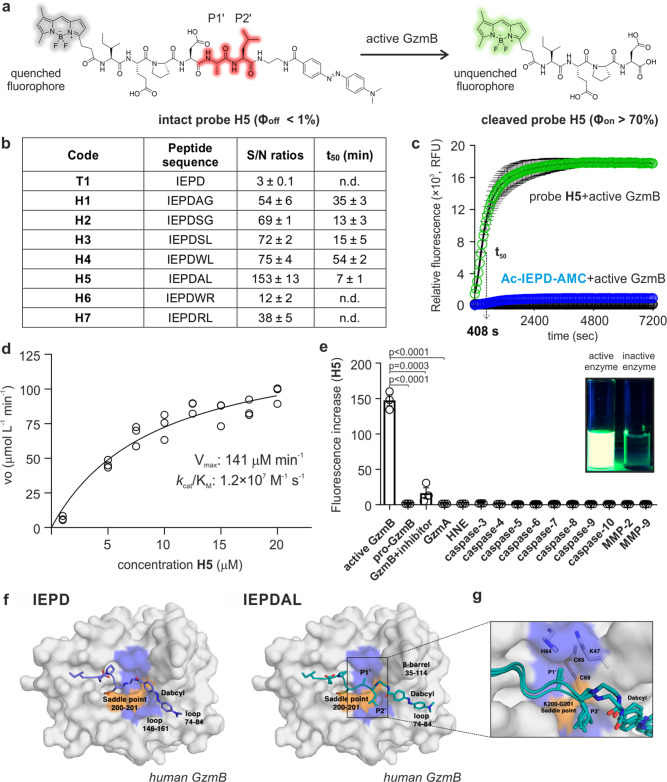
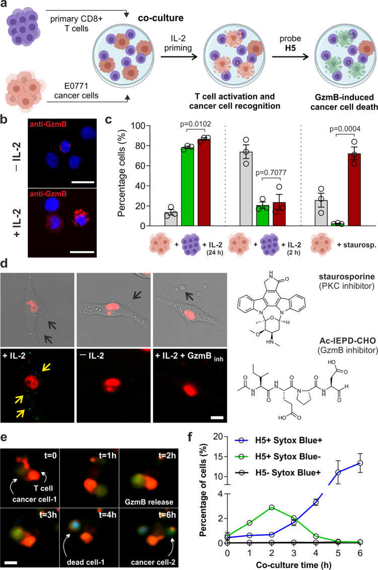
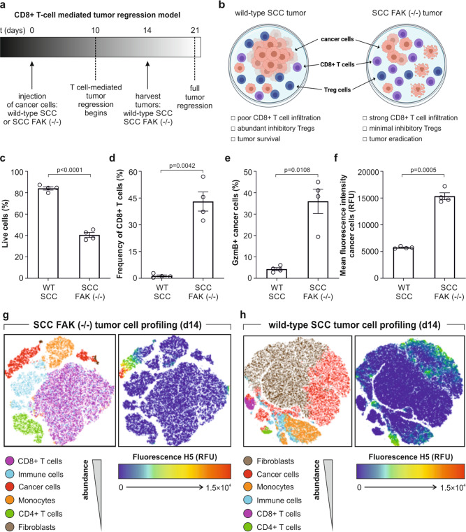
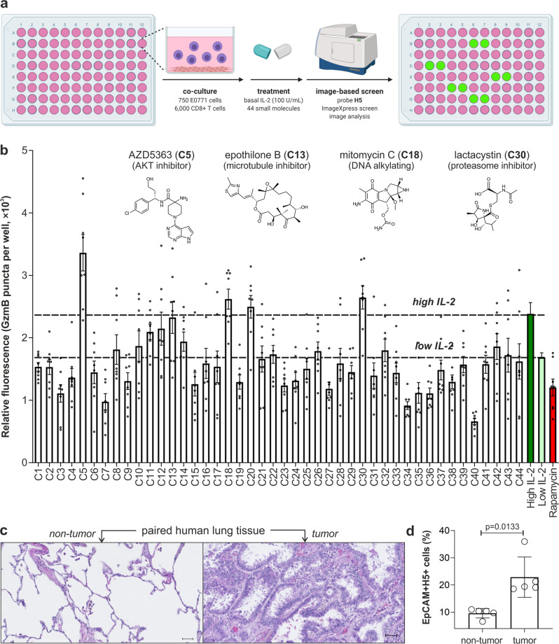
Immunotherapy promotes the attack of cancer cells by the immune system; however, it is difficult to detect early responses before changes in tumor size occur. Here, we report the rational design of a fluorogenic peptide able to detect picomolar concentrations of active granzyme B as a biomarker of immune-mediated anticancer action. Through a series of chemical iterations and molecular dynamics simulations, we synthesize a library of FRET peptides and identify probe H5 with an optimal fit into granzyme B. We demonstrate that probe H5 enables the real-time detection of T cell-mediated anticancer activity in mouse tumors and in tumors from lung cancer patients. Furthermore, we show image-based phenotypic screens, which reveal that the AKT kinase inhibitor AZD5363 shows immune-mediated anticancer activity. The reactivity of probe H5 may enable the monitoring of early responses to anticancer treatments using tissue biopsies.
© 2022. The Author(s).
Conflict of interest statement
The University of Edinburgh has filed a patent covering some of the technology described in this manuscript. J.M. is a current member of the Scientific Advisory Board of Cresset. The remaining authors declare no competing interests.
Figures
Similar articles
-
Fluorogenic Granzyme A Substrates Enable Real-Time Imaging of Adaptive Immune Cell Activity.Angew Chem Int Ed Engl. 2023 Feb 13;62(8):e202216142. doi: 10.1002/anie.202216142. Epub 2023 Jan 16. Angew Chem Int Ed Engl. 2023. PMID: 36562327 Free PMC article.
-
Granzyme B PET Imaging as a Predictive Biomarker of Immunotherapy Response.Cancer Res. 2017 May 1;77(9):2318-2327. doi: 10.1158/0008-5472.CAN-16-3346. Cancer Res. 2017. PMID: 28461564 Free PMC article.
-
In vivo bioluminescence imaging of granzyme B activity in tumor response to cancer immunotherapy.Cell Chem Biol. 2022 Oct 20;29(10):1556-1567.e6. doi: 10.1016/j.chembiol.2022.08.006. Epub 2022 Sep 13. Cell Chem Biol. 2022. PMID: 36103874 Free PMC article.
-
Imaging and monitoring of granzyme B in the immune response.Wiley Interdiscip Rev Nanomed Nanobiotechnol. 2024 Jan-Feb;16(1):e1928. doi: 10.1002/wnan.1928. Epub 2023 Sep 15. Wiley Interdiscip Rev Nanomed Nanobiotechnol. 2024. PMID: 37715320 Review.
-
Recent Advances in Lung Cancer Immunotherapy: Input of T-Cell Epitopes Associated With Impaired Peptide Processing.Front Immunol. 2019 Jul 3;10:1505. doi: 10.3389/fimmu.2019.01505. eCollection 2019. Front Immunol. 2019. PMID: 31333652 Free PMC article. Review.
Cited by
-
Fluorogenic Reactions in Chemical Biology: Seeing Chemistry in Cells.Chem Biomed Imaging. 2023 May 30;1(7):590-619. doi: 10.1021/cbmi.3c00029. eCollection 2023 Oct 23. Chem Biomed Imaging. 2023. PMID: 39474135 Free PMC article. Review.
-
Nonperturbative Fluorogenic Labeling of Immunophilins Enables the Wash-free Detection of Immunosuppressants.ACS Cent Sci. 2024 Mar 18;10(5):969-977. doi: 10.1021/acscentsci.3c01590. eCollection 2024 May 22. ACS Cent Sci. 2024. PMID: 38799658 Free PMC article.
-
Effect of refined management in operating room nursing on surgical efficiency and nursing satisfaction during laparoscopic radical resection of colon cancer.Am J Transl Res. 2024 Feb 15;16(2):506-514. doi: 10.62347/JUVO8905. eCollection 2024. Am J Transl Res. 2024. PMID: 38463602 Free PMC article.
-
Fluorogenic Granzyme A Substrates Enable Real-Time Imaging of Adaptive Immune Cell Activity.Angew Chem Int Ed Engl. 2023 Feb 13;62(8):e202216142. doi: 10.1002/anie.202216142. Epub 2023 Jan 16. Angew Chem Int Ed Engl. 2023. PMID: 36562327 Free PMC article.
-
Chemo-Click: Receptor-Controlled and Bioorthogonal Chemokine Ligation for Real-Time Imaging of Drug-Resistant Leukemic B Cells.J Am Chem Soc. 2024 Nov 6;146(44):30565-30572. doi: 10.1021/jacs.4c12035. Epub 2024 Oct 23. J Am Chem Soc. 2024. PMID: 39441736 Free PMC article.
References
Publication types
MeSH terms
Substances
Grants and funding
LinkOut - more resources
Full Text Sources
Other Literature Sources
Medical
