. 2022 May 2;13(1):2378.
doi: 10.1038/s41467-022-29950-w.

Unique binding pattern for a lineage of human antibodies with broad reactivity against influenza A virus

Affiliations

Unique binding pattern for a lineage of human antibodies with broad reactivity against influenza A virus

Xiaoyu Sun et al. Nat Commun. .

Abstract

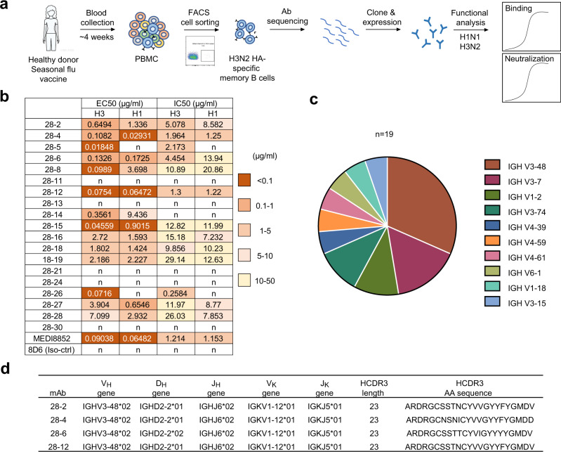
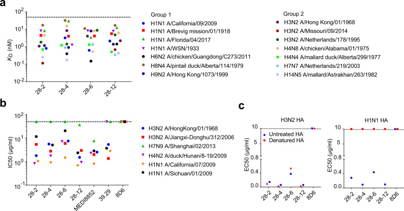
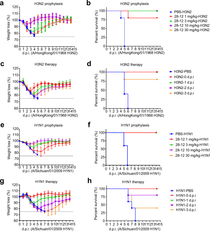
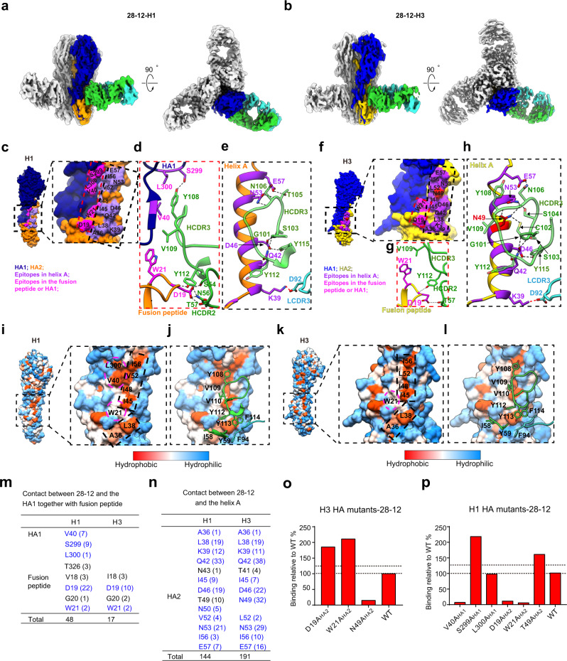
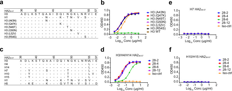
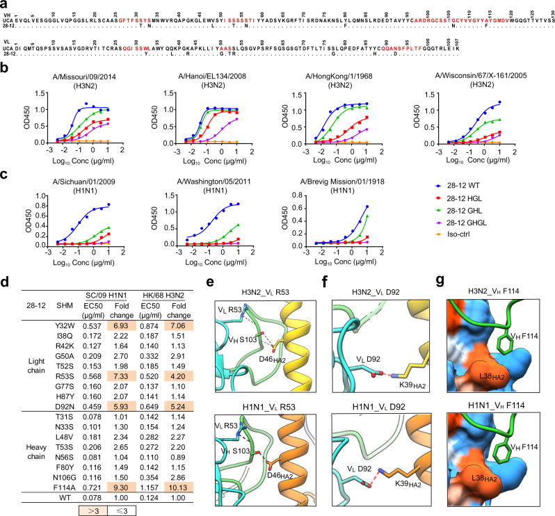
Most structurally characterized broadly neutralizing antibodies (bnAbs) against influenza A viruses (IAVs) target the conserved conformational epitopes of hemagglutinin (HA). Here, we report a lineage of naturally occurring human antibodies sharing the same germline gene, VH3-48/VK1-12. These antibodies broadly neutralize the major circulating strains of IAV in vitro and in vivo mainly by binding a contiguous epitope of H3N2 HA, but a conformational epitope of H1N1 HA, respectively. Our structural and functional studies of antibody 28-12 revealed that the continuous amino acids in helix A, particularly N49HA2 of H3 HA, are critical to determine the binding feature with 28-12. In contrast, the conformational epitope feature is dependent on the discontinuous segments involving helix A, the fusion peptide, and several HA1 residues within H1N1 HA. We report that this antibody was initially selected by H3 (group 2) viruses and evolved via somatic hypermutation to enhance the reactivity to H3 and acquire cross-neutralization to H1 (group 1) virus. These findings enrich our understanding of different antigenic determinants of heterosubtypic influenza viruses for the recognition of bnAbs and provide a reference for the design of influenza vaccines and more effective antiviral drugs.


Conflict of interest statement

B.S., X.S., X.L., and Z.L. are inventors on the patent application of antibody 28-12. The other authors declare no competing interests.

Figures

Fig. 1
Fig. 1. A lineage of antibodies with cross-reactivity to H3N2 and H1N1 subtypes were identified.
a Schematic of isolation of influenza HA specific antibodies from an individual vaccinated with trivalent inactivated influenza vaccine. The isolated antibodies were tested for dose-dependent binding and neutralizing activities against group 2 HK/68 H3N2 and group 1 SC/09 H1N1 (b). 8D6, an unrelated HCV antibody was included as a negative control. The EC50 and IC50 values are shown into heatmap. n, not binding or not neutralizing. c Distribution of V gene families in heavy chains of all distinct clones (n = 19). d Summary of the germline (V-D-J for heavy chain and V-J for light chain) and HCDR3 of VH3-48/VK1-12 lineage antibodies.
Fig. 2
Fig. 2. VH3-48/VK1-12 lineage antibodies broadly bind and neutralize multiple influenza subtypes.
a Binding affinity (KD) of each mAb to HA proteins from a panel of group 1 and group 2 influenza A isolates as measured by BLI. Dashed line indicates 50 nM. b In vitro neutralizing activity of each mAb against multiple strains of H1, H3, H4, and H7 subtype IAVs, shown as IC50 values. MEDI8852 and 39.29 were included as positive controls while 8D6 was the negative control. Dashed line indicates 50 μg/ml. The hosts of non-human infecting IAV strains have been noted in the full name of IAV strains, while the other IAV strains without host annotation are human infecting. c ELISA binding EC50 values of each mAb to denatured or untreated HAs of H3N2 and H1N1. Representative data are shown from two independent experiments.
Fig. 3
Fig. 3. Prophylactic and therapeutic efficacy of 28-12 in mice.
a, b Prophylactic efficacy of 28-12 against a lethal challenge with 68/HK H3. Mice were treated with 30, 10, 3 or 1 mg/kg 28-12 or PBS 24 h before intranasal inoculation with the influenza virus. The weight loss and survival data were collected daily for 14 days after inoculation (day 0). n = 5. c, d Therapeutic efficacy of 28-12 against a lethal challenge with 68/HK H3. Mice were treated with PBS buffer (at day 0) or 25 mg/kg 28-12 immediately after or 1, 2, or 3 days after intranasal inoculation with influenza virus. The weight loss and survival data were collected daily for 14 days after inoculation with viruses (day 0). n = 5. e, f Prophylactic efficacy of 28-12 against a lethal challenge with SC/09 H1. n = 5. g, h Therapeutic efficacy of 28-12 (25 mg/kg) against a lethal challenge with SC/09 H1. n = 5. Error bars represent the mean ± S.D (a, c, e, g). All data show a representative experiment from two independent experiments.
Fig. 4
Fig. 4. Cryo-EM structures of 28-12-H3 and 28-12-H1 and distinct recognition patterns of 28-12 Fab to H3 and H1 HAs.
a, b Cryo-EM maps of the 28-12-H1 (a) and 28-12-H3 (b) complexes. One HA protomer and the cognate 28-12 Fab fragment are highlighted in color, and the other two copies in the trimer are colored gray. H1 HA1/HA2 and H3 HA1/HA2 are colored in navy blue, orange, blue, and gold, respectively. The heavy and light chains of 28-12 Fab are colored in lime green and turquoise, respectively. This color scheme was followed throughout. c, f Epitopes of the 28-12 Fab on H1 (c) and H3 (f), respectively. The epitopes in helix A (indicated by black dashed box) and fusion peptide/HA1 (indicated by red dashed box) are colored in purple and magenta, respectively. H3 residue N49HA2 is specifically showed in red. d, g Hydrogen bonds between 28-12 Fab and the fusion peptide/HA1 of H1 (d) or H3 (g). e, h Hydrogen bonds formed between helix A of H1 (e) or H3 (h) and 28-12 Fab. The salt bridge and hydrogen bond are shown as red and black dashed lines, respectively. i, k Hydrophobic epitopes in the H1 (i) and H3 (k) HAs. The hydrophobic epitopes are classified into two parts, helix A in the dashed black box and the fusion peptide and HA1 residues in the dashed magenta circle. j, l The Fab residues involved in hydrophobic contacts with H1 (j) and H3 (l) are shown with side chains. m The number of atom-to-atom contacts between 28-12 Fab and the fusion peptide/HA1 residues. n The number of atom-to-atom contacts between 28-12 Fab and the helix A. The epitopes involved in the hydrophobic interaction, hydrogen bond, and salt bridge are colored in blue. o, p 28-12 binding to H1 and H3 HA mutants with alanine substitutions. Each value is calculated as the binding ratio relative to that of the WT HA (%). The two black dotted lines represent 100 and 125% relative binding to the WT data, respectively. The mean values of duplicates are shown from two independent experiments (o, p).
Fig. 5
Fig. 5. N49HA2 is critical for the recognition of H3 epitope features by VH3-48/VK1-12 lineage antibodies.
a Sequence alignment of the conserved epitope 36-57HA2 of H1 and H3. b Dose-dependent binding of 28-12 to divergent mutants of H3 peptide 36-57HA2 compared with the wild-type peptide. c Sequence alignment of 36-57HA2 within multiple subtypes. H3/H4/H14, but not other subtypes have residue N in position 49HA2. df Dose-dependent reactivity of the VH3-48/VK1-12 lineage antibodies to peptide 36-57HA2 of multiple subtypes. 8D6 was included as the isotype control. Data are presented as mean values of duplicates (b, d, e, f). Representative data are shown from two independent experiments (b, d, e, f).
Fig. 6
Fig. 6. Somatic hypermutations of 28-12.
a Sequence alignment of 28-12 and its UCA. Dots indicate identical residues. CDR regions according to IMGT are shown in red. b, c The reactivity of 28-12 germline variants to H3N2 (b) and H1N1 HAs (c). GHGL, VH germline paired with VL germline. HGL, 28-12 VH paired with its VL germline. GHL, 28-12 VL paired with its VH germline. d Heatmap showing the EC50 values of 28-12 variants with single somatic mutations to H3N2 and H1N1 HAs. e For both 28-12-H3 and 28-12-H1 complexes, VL R53 forms hydrogen bonds with the main chain of VH S103 in HCDR3, which also contacts the residue D46HA2 through hydrogen bonds. f For both 28-12-H3 and 28-12-H1 complexes, VL D92 of 28-12 forms salt bridges with K39HA2 of helix A. g VH F114 inserts into a hydrophobic groove formed by L38HA2 of both H3N2 and H1N1 HAs. The colors of HA1 and HA2 and the heavy chain and light chain of 28-12 are shown as in Fig. 4. The salt bridge and hydrogen bond are shown as red and black dashed lines, respectively. Data are presented as mean values of duplicates (b, c). Representative data are shown from two independent experiments (b, c).

Similar articles

Cited by

References

    1. Coughlan L, Palese P. Overcoming barriers in the path to a universal influenza virus vaccine. Cell Host Microbe. 2018;24:18–24. doi: 10.1016/j.chom.2018.06.016. - DOI - PubMed
    1. Medina RA, Garcia-Sastre A. Influenza A viruses: new research developments. Nat. Rev. Microbiol. 2011;9:590–603. doi: 10.1038/nrmicro2613. - DOI - PMC - PubMed
    1. Krammer F. The human antibody response to influenza A virus infection and vaccination. Nat. Rev. Immunol. 2019;19:383–397. doi: 10.1038/s41577-019-0143-6. - DOI - PubMed
    1. Nobusawa E, et al. Comparison of complete amino acid sequences and receptor-binding properties among 13 serotypes of hemagglutinins of influenza A viruses. Virology. 1991;182:475–485. doi: 10.1016/0042-6822(91)90588-3. - DOI - PubMed
    1. Meissner HC, et al. Immunoprophylaxis with palivizumab, a humanized respiratory syncytial virus monoclonal antibody, for prevention of respiratory syncytial virus infection in high risk infants: a consensus opinion. Pediatr. Infect. Dis. J. 1999;18:223–231. doi: 10.1097/00006454-199903000-00004. - DOI - PubMed

Publication types