Review
. 2022 May 2;8(1):239.
doi: 10.1038/s41420-022-01034-0.

The pathological role of damaged organelles in renal tubular epithelial cells in the progression of acute kidney injury

Affiliations
Review

The pathological role of damaged organelles in renal tubular epithelial cells in the progression of acute kidney injury

Zixian Li et al. Cell Death Discov. .

Abstract

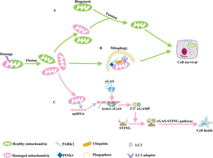
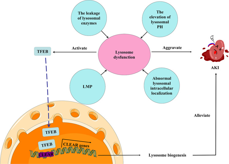
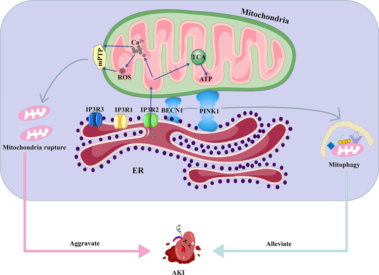
Acute kidney injury (AKI) is a common clinical condition associated with high morbidity and mortality. The pathogenesis of AKI has not been fully elucidated, with a lack of effective treatment. Renal tubular epithelial cells (TECs) play an important role in AKI, and their damage and repair largely determine the progression and prognosis of AKI. In recent decades, it has been found that the mitochondria, endoplasmic reticulum (ER), lysosomes, and other organelles in TECs are damaged to varying degrees in AKI, and that they can influence each other through various signaling mechanisms that affect the recovery of TECs. However, the association between these multifaceted signaling platforms, particularly between mitochondria and lysosomes during AKI remains unclear. This review summarizes the specific pathophysiological mechanisms of the main TECs organelles in the context of AKI, particularly the potential interactions among them, in order to provide insights into possible novel treatment strategies.


Conflict of interest statement

The authors declare no competing interests.

Figures

Fig. 1
Fig. 1. Mitochondrial dynamics during AKI.
Mitochondria undergo the dynamic balance of fission and fusion to maintain normal mitochondrial function. A In AKI, damaged mitochondria could also occur fission. Then biogenesis and fusion help mix partially damaged mitochondria to produce healthy mitochondria. B Additionally, mitophagy are involved in maintaining mitochondrial function and cell homeostasis through degrading damaged or incomplete mitochondria. C However, once the dynamics of mitochondria are out of balance, mtDNA is released from damaged mitochondria to the cytoplasm. Subsequently, the cGAS-STING pathway is activated and cell apoptosis occurs. AKI Acute kidney injury, mtDNA Mitochondrial DNA.
Fig. 2
Fig. 2. Lysosome dysfunction and TFEB-mediated lysosome biogenesis during AKI.
The leakage of lysosomal enzymes, abnormal lysosomal intracellular localization, the elevation of lysosomal pH and LMP could cause lysosome dysfunction, thereby aggravating renal injury during AKI. These pathological conditions induce the nuclear localization of TFEB. TFEB activates CLEAR network genes to control lysosomal biogenesis, which could alleviate AKI. LMP Lysosomal membrane permeabilization, TFEB Transcription factor EB, CLEAR Coordinated lysosomal expression and regulation.
Fig. 3
Fig. 3. ERS after AKI.
Major ER transmembrane proteins mainly include IRE1, PERK and ATF6. Under normal physiological conditions, they are all bound to Bip / GRP78 to form a stable complex. Under various acute pathological stimuli such as hypoxia, the reduction of ATP, nutrient deficiency, and an increase in ROS, tubular cells often undergo ERS. The three ER transmembrane proteins disassociate with Bip to collectively execute the UPR. The UPR pathway consists of the adaptive UPR pathway which could maintain cell function and the apoptotic pathway which could cause cell death. ER endoplasmic reticulum, ERS endoplasmic reticulum stress, IRE1 inositol-requiring protein 1, PERK protein kinase RNA-like ER kinase, ATF6 activating transcription factor 6, UPR unfolded protein response.
Fig. 4
Fig. 4. Ca2+ balance mediated by MAMs.
IP3R is the main Ca2+ release channel located at the ER membrane, designated as type 1, type 2, and type 3. Mitochondrial Ca2+ overload by MAMs boosts ATP production by stimulating TCA cycles and produces a mass of ROS. The opening of the mPTP results in mitochondria rupture, which could aggravate AKI. However, BECN1/PINK1 expression enhances MAMs formation and promotes mitophagy to alleviate AKI. MAMs mitochondria-associated ER membranes, IP3R Inositol 1,4,5-trisphosphate receptor, mPTP mitochondrial permeability transition pore, BECN1 Recombinant Beclin 1, PINK1 PTEN-induced kinase 1.
Fig. 5
Fig. 5. The regulation of mitochondria-lysosome membrane contact.
A Lysosome and mitochondria could establish crosstalk by mitochondria-lysosome membrane contact. B RAB7 GTPase-activating protein TBC1D15 is recruited to mitochondria by FIS1, thereby mediating mitochondria-lysosome membrane contact tethering. C Lysosomal dynamics are regulated by the RAB7 GTPase, which cycles from an active GTP-bound state to an inactive GDP-bound state. D AKI leads to mitochondria dysfunction, and the mitochondria produce a large amount of ROS. Meanwhile, the lysosomal channel TRPML1, which is the ROS sensor localized on the lysosomal membrane, is triggered, increasing mitochondrial Ca2+. The contacts become unstable and untether. In addition, lysosomal damage facilitates Ca2+ efflux through TRPML1, which is essential for TFEB activation to induce mitophagy. FIS1 Mitochondrial fission 1, TRPML1 Transient receptor potential mucolipin 1.

Similar articles

Cited by

References

    1. Al-Jaghbeer M, Dealmeida D, Bilderback A, Ambrosino R, Kellum JA. Clinical decision support for in-hospital AKI. J Am Soc Nephrology: JASN. 2018;29:654–60. doi: 10.1681/ASN.2017070765. - DOI - PMC - PubMed
    1. Ronco C, Bellomo R, Kellum JA. Acute kidney injury. Lancet (Lond, Engl) 2019;394:1949–64. doi: 10.1016/S0140-6736(19)32563-2. - DOI - PubMed
    1. Schefold JC, Filippatos G, Hasenfuss G, Anker SD, von Haehling S. Heart failure and kidney dysfunction: Epidemiology, mechanisms and management. Nat Rev Nephrol. 2016;12:610–23. doi: 10.1038/nrneph.2016.113. - DOI - PubMed
    1. Basile DP, Anderson MD, Sutton TA. Pathophysiology of acute kidney injury. Compr Physiol. 2012;2:1303–53. doi: 10.1002/cphy.c110041. - DOI - PMC - PubMed
    1. Lewington AJ, Cerdá J, Mehta RL. Raising awareness of acute kidney injury: A global perspective of a silent killer. Kidney Int. 2013;84:457–67. doi: 10.1038/ki.2013.153. - DOI - PMC - PubMed