. 2022 May 2;12(1):7108.
doi: 10.1038/s41598-022-11043-9.

Role of cathepsin K in the expression of mechanical hypersensitivity following intra-plantar inflammation

Affiliations

Role of cathepsin K in the expression of mechanical hypersensitivity following intra-plantar inflammation

Maha Paracha et al. Sci Rep. .

Abstract

Persistent/chronic inflammatory pain involves multiple pathophysiological mechanisms and is far more complex than acute/momentary pain. Current therapeutics for chronic inflammatory pain are often not effective because the etiology responsible for the pain is not addressed by traditional pharmacological treatments. Cathepsin K is a cysteine protease that has mostly been studied in the context of bone and joint disorders. Previous work by others has shown that inhibition of cathepsin K activity reduces osteoarthritis-associated nociception in joints. However, the role of cathepsin K in cutaneous inflammation is understudied. We assessed the effectiveness of genetic deletion or pharmacological inhibition of cathepsin K in male mice on the expression of nocifensive behaviors after formalin injection or mechanical and thermal hypersensitivity after injection of complete Freund's adjuvant (CFA) into the mouse hind paw. Our data demonstrate that cathepsin K knockout mice (Ctsk-/-) have a reduction in nocifensive behaviors in the formalin test. In addition, Ctsk-/- do not develop mechanical hypersensitivity after CFA injection for up to 7 days. Moreover, we found that inhibition of cathepsin K reduced mechanical hypersensitivity after CFA injection and mRNA levels, protein levels, and cathepsin K activity levels were elevated after CFA injection. Based upon our data, cathepsin K is indicated to play a role in the expression of chemically-induced cutaneous hypersensitivity, as Ctsk-/- mice do not develop mechanical hypersensitivity and show a reduction in nocifensive behaviors. Further research is needed to determine whether attenuating cathepsin K activity may generate a clinically relevant therapeutic.


Conflict of interest statement

The authors declare no competing interests.

Figures
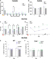
Figure 1
Figure 1
Cathepsin K knockout mice do not develop hypersensitivity to mechanical or thermal stimuli and have an attenuation in chemically induced nocifensive behaviors. (A) Formalin test, cathepsin K knockout (Ctsk−/−) mice have a reduction in nocifensive behaviors associated with the longer-lasting, more persistent phase of the formalin test compared to wild type (WT) mice. Orange triangle indicates the time of formalin injection. (B) General locomotor activity between WT and Ctsk−/− mice. (C,D) Compared to the WT mice, Ctsk−/− mice do not show mechanical hypersensitivity after complete Freund’s adjuvant (CFA) injection, shown as scatter plot overlayed with a bar graph representing the median; (D) line graph of the same data. Red triangle indicates the time of CFA injection. (E) CFA-injected WT mice show a trend towards thermal hypersensitivity after CFA injection, which is not seen in the CFA-injected Ctsk−/− mice (F). Data are represented at scatter plots of individual mice with bar graphs overlayed indicating the median value. *p < 0.05 multiple Mann–Whitney test between WT and Ctsk−/− mice for each time point.
Figure 2
Figure 2
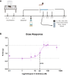
Dose–response curve of cathepsin K inhibitor in mice. (A) Timeline for the dose–response curve. Nine different concentrations of cathepsin K inhibitor were selected. Each group of WT mice was given an injection of one of the nine different concentrations of the cathepsin K inhibitor on day 1 post-CFA (complete Freund’s adjuvant) injection. Their response to each concentration of the inhibitor in terms of force (g) needed to induce paw withdrawal was recorded and these responses were used to plot a dose–response curve. (B) The calculated IC50 of the inhibitor is 23.26 nM, the top of the response curve lies at 1.51 g and the bottom of the response curve lies at 0.61 g. Of the doses tested, the inhibitor starts to show an effect at 5 nM and the effect peaks at 50 nM. Data are represented as mean ± SEM.
Figure 3
Figure 3
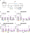
Cathepsin K inhibitor (50 nM) reduces mechanical hypersensitivity in mice. (A) Timeline for testing mechanical hypersensitivity. (B) There is no effect of the 50 nM cKi when given in mice with saline injection. Blue and yellow triangle indicates the time of saline and cKi injection respectively. (C) Whereas a single injection of 50 nM cKi (yellow triangle) when given before testing on day 1 post-CFA (complete Freund’s adjuvant) injection (red triangle) results in higher mechanical threshold as compared to vehicle (10% DMSO in saline) injection. (D) During the developmental phase of chronic inflammatory pain (i.e., on day 1 and day 2 post-CFA injection), subsequent injections of 50 nM cKi can alleviate hypersensitivity compared to vehicle injection. (E) Similarly, during the maintenance phase of persistent inflammatory pain (i.e., on day 7 post-CFA injection) 50 nM cKi injection reduces mechanical hypersensitivity when compared to vehicle injection. Data are represented at scatter plots of individual mice with bar graphs overlayed indicating the median value. *p < 0.05 Mann–Whitney test between cKi and vehicle treatment for each day.
Figure 4
Figure 4
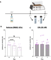
Cathepsin K inhibitor (50 nM) reduces thermal hypersensitivity in mice. (A) Timeline for testing thermal hypersensitivity. (B) Vehicle-injected mice have a significantly lower thermal latency than day 0 (baseline) on day 7 post-CFA injection. (C) 50 nM cKi-injected mice show no significant change in thermal latency than day 0 (baseline) post-CFA injection. Data are represented at scatter plots of individual mice with bar graphs overlayed indicating the median value. *p < 0.05 Mann–Whitney test between time points for each treatment.
Figure 5
Figure 5
Cathepsin K levels rise post-inflammation in mice. (A) Timeline for testing mRNA, protein levels and protein activity. (B) There is an increase in cathepsin K mRNA levels in the hind paw after CFA injection. (C) Increased cathepsin K protein (active-form) in hind paw after CFA injection. (D) In addition, there is an increase in cathepsin K activity in the hind paw after CFA injection. (E) Representative western blot of cathepsin K and housekeeping protein GAPDH after CFA injection. Black arrow indicates the molecular weight of detected protein. CFA and Saline treated samples are represented as C and S, respectively. Data are represented at scatter plots of individual mice with bar graphs overlayed indicating the median value. *p < 0.05 Mann–Whitney test between saline and CFA treatment on day 1 and day 7.

Similar articles

References

    1. Woolf CJ. What is this thing called pain? J. Clin. Invest. 2010;120:3742–3744. doi: 10.1172/JCI45178. - DOI - PMC - PubMed
    1. Tansley SN, Wong C, Uttam S, Mogil JS, Khoutorsky A. Translation regulation in the spinal dorsal horn—a key mechanism for development of chronic pain. Neurobiol. Pain. 2018;4:20–26. doi: 10.1016/j.ynpai.2018.03.003. - DOI - PMC - PubMed
    1. Cenac N. In: Proteases and Their Receptors in Inflammation Progress in Inflammation Research. Chignard N, Vergnolle M, editors. Springer; 2011. pp. 253–274.
    1. Feizerfan A, Sheh G. Transition from acute to chronic pain. Contin. Educ. Anaesth. Crit. Care Pain. 2015;15:98–102. doi: 10.1093/bjaceaccp/mku044. - DOI
    1. Matsuda M, Huh Y, Ji RR. Roles of inflammation, neurogenic inflammation, and neuroinflammation in pain. J. Anesth. 2019;33:131–139. doi: 10.1007/s00540-018-2579-4. - DOI - PMC - PubMed

Publication types