. 2022 Jun;39(6):1261-1276.
doi: 10.1007/s10815-022-02495-z. Epub 2022 May 1.

Zinc transporters ZIPT-2.4 and ZIPT-15 are required for normal C. elegans fecundity

Affiliations

Zinc transporters ZIPT-2.4 and ZIPT-15 are required for normal C. elegans fecundity

Aaron C Sue et al. J Assist Reprod Genet. 2022 Jun.

Abstract

Purpose: The requirement of zinc for the development and maturation of germ lines and reproductive systems is deeply conserved across evolution. The nematode Caenorhabditis elegans offers a tractable platform to study the complex system of distributing zinc to the germ line. We investigated several zinc importers to investigate how zinc transporters play a role in the reproductive system in nematodes, as well as establish a platform to study zinc transporter biology in germline and reproductive development.

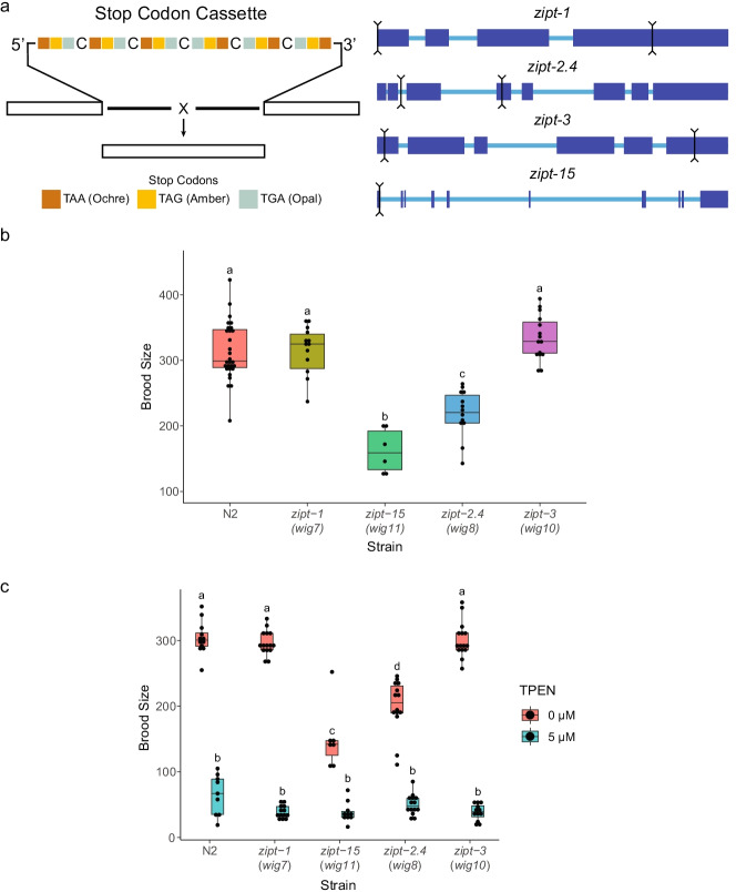
Methods: Previous high throughput transcriptional datasets as well as phylogenetic analysis identified several putative zinc transporters that have a function in reproduction in worms. Phenotypic analysis of CRISPR-generated knockouts and tags included characterization of offspring output, gonad development, and protein localization. Light and immunofluorescence microscopy allowed for visualization of physiological and molecular effects of zinc transporter mutations.

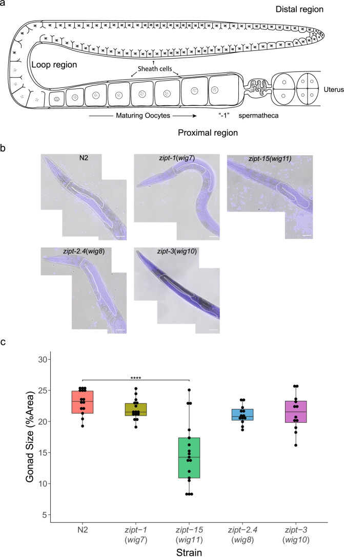
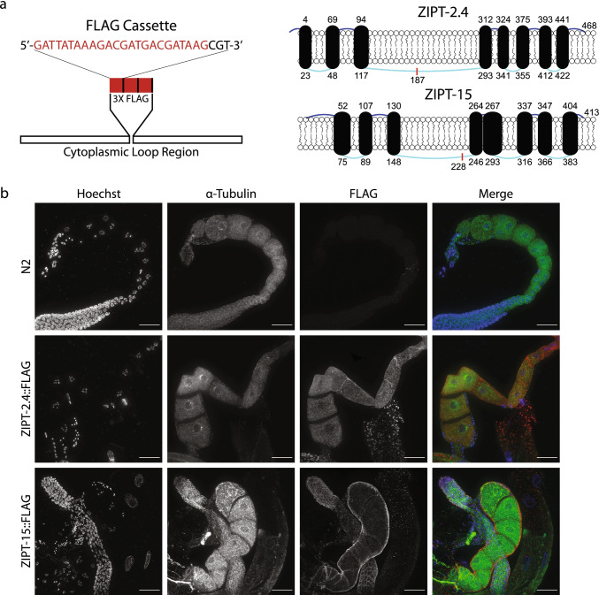
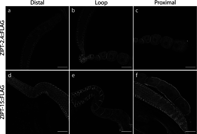
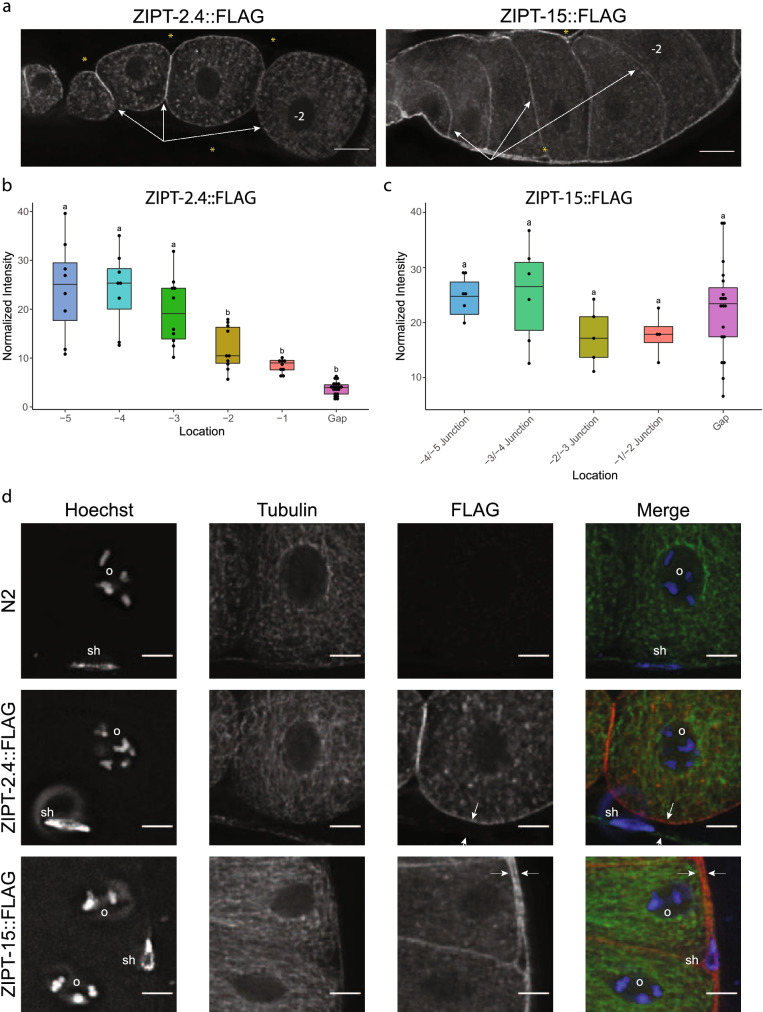
Results: Disruption of two zinc transporters, ZIPT-2.4 and ZIPT-15, was shown to lead to defects in reproductive output. A mutation in zipt-2.4 has subtle effects on reproduction, while a mutation in zipt-15 has a clear impact on gonad and germline development that translates into a more pronounced defect in fecundity. Both transporters have germline expression, as well as additional expression in other cell types.

Conclusions: Two ZIP-family zinc transporter orthologs of human ZIP6/10 and ZIP1/2/3 proteins are important for full reproductive fecundity and participate in development of the gonad. Notably, these zinc transporters are present in gut and reproductive tissues in addition to the germ line, consistent with a complex zinc trafficking network important for reproductive success.

Keywords: Caenorhabditis elegans; Fecundity; Germline development; Germline gene expression; Zinc transporter.


Conflict of interest statement

The authors declare no competing interests.

Figures

Fig. 1
Fig. 1
Identification of brood size defects in zinc transporter mutants. a Cartoon depicting the stop codon array used to generate each zinc transporter mutant. Sets of stop codons were designed in every reading frame, and a perfect insertion of the cassette will also introduce a frameshift. The exons of each gene are depicted in the larger blue boxes. The black lines indicate the 5’ and 3’ cuts made to replace the intervening sequence with the stop codon array. For zipt-15, only one cut was made, and the cassette was inserted at this point. Box plots of the brood sizes of each zinc transporter mutant for the first 5 days of reproductive adulthood (b) and challenged with either 0 or 5 μM TPEN (c). Each plot represents 3 biological replicates with 5 technical replicates per experiment. Data for worms that died before the experiment completed were discarded. Box plots labeled with the same letter are statistically indistinguishable (p > 0.05) as determined by the post hoc Tukey HSD test
Fig. 2
Fig. 2
Examination of the gonad architecture of zinc transporter mutants. a Cartoon depicting the general structure of a gonad arm in C. elegans. The distal region contains the mitotic niche, as well as early meiotic cells. Except for the most distal region of the gonad, the germ line is encased by 5 sets of cells called the sheath cells. As the cells enter the loop region, their membrane completely encloses to form oocytes. As these oocytes enter the proximal region, they become prepared for fertilization, arresting at Prophase I of meiosis. The most mature oocyte is referred to as “-1.” Oocytes are processively pushed through the spermatheca, which houses the sperm, and subsequently ejected into a uterus that is shared between both gonad arms. b Representative slices of stitched images of N2, zipt-1, zipt-15, zipt-2.4, and zipt-3 mutants stained with Hoechst. The white lines represent a trace of the gonad arm. Scale bar represents 50 μm. c Box plots of the relative gonad arm size of each zinc transporter mutant. Each plot represents at least 10 technical replicates. Significance values shown as a Tukey HSD test relative to control. ****p < 1E-6
Fig. 3
Fig. 3
Generation of FLAG-tagged zinc transporter strains. a Cartoon representing the FLAG cassette used to tag ZIPT-2.4 and ZIPT-15. Each FLAG array (red) was separated by one arginine (black) and inserted in frame into the predicted cytoplasmic loop regions. The cylinders represent the transmembrane domains, which are connected by extracellular (dark blue) and intracellular (light blue) strands. Each number indicates the beginning and ending amino acid of the TM domains, and the lengths of the TM domains and loops are scaled to their size relative to the whole protein, with the exception of the N terminal strand of ZIPT-2.4 which was increased for clarity. The red line indicates the location of the FLAG insertion. b Representative maximum projections of wild-type (N2) and FLAG-tagged strains probed with FLAG (red) and tubulin (green) antibodies, as well as Hoechst to label DNA (blue). All images were taken under the same exposure conditions, and all images are displayed using the same minimum and maximum intensity values. Scale bar represents 25 μm
Fig. 4
Fig. 4
ZIPT-2.4 and ZIPT-15 are expressed in multiple regions of the gonad. Representative slices of the distal (a, d), loop (b, e), and proximal (c, f) regions in the gonads of the ZIPT-2.4::FLAG (top) and ZIPT-15::FLAG (bottom) strains, stained with an anti-FLAG antibody. All images were exposed and processed under equivalent conditions. Scale bar represents 25 μm
Fig. 5
Fig. 5
ZIPT-2.4::FLAG and ZIPT-15::FLAG have distinct expression patterns in the gonad. a Representative slices of ZIPT-2.4::FLAG and ZIPT-15::FLAG signal. In the images shown, oocyte maturity (age) is increasing from left to right, with the -2 oocyte labeled in each image. White arrows indicate oocyte membrane signal, which decreases from left to right. Yellow asterisks indicate either locations where sheath cell FLAG signal is weak or absent (ZIPT-2.4) or strong (ZIPT-15). b, c Box plots of normalized fluorescence intensity values of ZIPT-2.4::FLAG (b) and ZIPT-15::FLAG (c) associated with oocyte membranes and non-oocyte membranes adjacent to the oocyte. Box plots labeled with the same letter are statistically indistinguishable (p > 0.05) as determined by the post hoc Tukey HSD test. d Representative Z slices of zoomed insets of N2, ZIPT-2.4::FLAG, and ZIPT-15::FLAG immunofluorescence images. Shown are FLAG (red), tubulin (green), and DNA (blue). Each image contains an oocyte nucleus (labeled “o”), as well as genetic material not associated with oocytes; nuclei putatively associated with sheath cells are labeled “sh.” The white arrows indicate two membranes, signifying another cell type contacting the oocyte, likely the sheath cell, as well as the outer membrane of the putative sheath cell. All images were exposed and processed under equivalent conditions. Scale bar represents 5 μm

Similar articles

Cited by

References

    1. Quesada-Candela C, Loose J, Ghazi A, Yanowitz JL. Molecular basis of reproductive senescence: insights from model organisms. J Assist Reprod Genet. 2021;38:17–32. doi: 10.1007/s10815-020-01959-4. - DOI - PMC - PubMed
    1. Gu L, et al. Metabolic control of oocyte development: linking maternal nutrition and reproductive outcomes. Cell Mol Life Sci. 2015;72:251–271. doi: 10.1007/s00018-014-1739-4. - DOI - PMC - PubMed
    1. Lin YJ, et al. Detrimental effect of maternal and post-weaning high-fat diet on the reproductive function in the adult female offspring rat: roles of insulin-like growth factor 2 and the ovarian circadian clock. J Assist Reprod Genet. 2017;34:817–826. doi: 10.1007/s10815-017-0915-5. - DOI - PMC - PubMed
    1. Howie GJ, Sloboda DM, Kamal T, Vickers MH. Maternal nutritional history predicts obesity in adult offspring independent of postnatal diet. J Physiol. 2009;587:905–915. doi: 10.1113/jphysiol.2008.163477. - DOI - PMC - PubMed
    1. Gaskins AJ, et al. Dietary folate and reproductive success among women undergoing assisted reproduction. Obstet Gynecol. 2014;124:801–809. doi: 10.1097/AOG.0000000000000477. - DOI - PMC - PubMed

MeSH terms

LinkOut - more resources