. 2022 Jul;59(7):4159-4178.
doi: 10.1007/s12035-022-02838-y. Epub 2022 Apr 30.

Treatment with IFB-088 Improves Neuropathy in CMT1A and CMT1B Mice

Affiliations

Treatment with IFB-088 Improves Neuropathy in CMT1A and CMT1B Mice

Yunhong Bai et al. Mol Neurobiol. 2022 Jul.

Abstract

Charcot-Marie-Tooth disease type 1A (CMT1A), caused by duplication of the peripheral myelin protein 22 (PMP22) gene, and CMT1B, caused by mutations in myelin protein zero (MPZ) gene, are the two most common forms of demyelinating CMT (CMT1), and no treatments are available for either. Prior studies of the MpzSer63del mouse model of CMT1B have demonstrated that protein misfolding, endoplasmic reticulum (ER) retention and activation of the unfolded protein response (UPR) contributed to the neuropathy. Heterozygous patients with an arginine to cysteine mutation in MPZ (MPZR98C) develop a severe infantile form of CMT1B which is modelled by MpzR98C/ + mice that also show ER stress and an activated UPR. C3-PMP22 mice are considered to effectively model CMT1A. Altered proteostasis, ER stress and activation of the UPR have been demonstrated in mice carrying Pmp22 mutations. To determine whether enabling the ER stress/UPR and readjusting protein homeostasis would effectively treat these models of CMT1B and CMT1A, we administered Sephin1/IFB-088/icerguestat, a UPR modulator which showed efficacy in the MpzS63del model of CMT1B, to heterozygous MpzR98C and C3-PMP22 mice. Mice were analysed by behavioural, neurophysiological, morphological and biochemical measures. Both MpzR98C/ + and C3-PMP22 mice improved in motor function and neurophysiology. Myelination, as demonstrated by g-ratios and myelin thickness, improved in CMT1B and CMT1A mice and markers of UPR activation returned towards wild-type values. Taken together, our results demonstrate the capability of IFB-088 to treat a second mouse model of CMT1B and a mouse model of CMT1A, the most common form of CMT. Given the recent benefits of IFB-088 treatment in amyotrophic lateral sclerosis and multiple sclerosis animal models, these data demonstrate its potential in managing UPR and ER stress for multiple mutations in CMT1 as well as in other neurodegenerative diseases. (Left panel) the accumulation of overexpressed PMP22 or misfolded mutant P0 in the Schwann cell endoplasmic reticulum (ER) leads to overwhelming of the degradative capacity, activation of ER-stress mechanisms, and myelination impairment. (Right panel) by prolonging eIF2α phosphorylation, IFB-088 reduces the amount of newly synthesized proteins entering the ER, allowing the protein quality control systems to better cope with the unfolded/misfolded protein and allowing myelination to progress.

Keywords: Charcot-Marie-Tooth; IFB-088/Sephin1/icerguestat; Neuropathy; Proteostasis; UPR.


Conflict of interest statement

P.G., P.M. and C.T. are full-time employees and stockholders of InFlectis BioScience. M.D. acts as a Scientific Advisory Board member and Consultant for InFlectis BioScience.

Figures
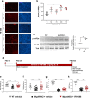
Fig. 1
Fig. 1
IFB-088 improves myelination in DRG explants, motor function and nerve conduction velocity from MpzR98C/ + mice. Dorsal root ganglia (DRG) were dissected from embryos (E13.5) of MpzR98C/ + mice. The myelination process was induced with ascorbic acid. After 2 weeks of treatment with vehicle or the indicated concentration of IFB-088, the DRGs were fixed and nuclei visualized by DAPI staining; myelin were visualized by immunostaining with myelin basic protein (MBP) antibody. a Representative pictures; scale bar 100 μm. b Number of myelinated internodes per field in MpzR98C/ + DRG explant cultures treated with vehicle or the indicated concentration of IFB-088 for 2 weeks. Mean ± SEM; n = 3 independent experiments. *P < 0.05 one-way ANOVA followed by Friedman’s test. c Representative western blot (WB) for P-eIF2α, total eIF2α and tubulin on sciatic nerve protein extracts from 1-month-old WT and MpzR98C/ + mice. The graph shows the densitometric quantification of the P-eIF2α/eIF2α ratio; *P < 0.05 by unpaired t-test. d Diagram of the treatment strategy. Thirty-day-old WT and MpzR98C/ + mice were orally administered with vehicle or IFB-088 1 mg/kg b.i.d. for 5 months. e Four limb grip strength max values average of 10 trials. Data were expressed in grams (g) as mean ± SEM; n = 15–26 mice per condition. (f) Rotarod analysis. Data are expressed in seconds (s) as mean ± SEM; n = 15–26 mice per condition. g Motor nerve conduction velocity (MNCV). Data are expressed in meters/second (m/s) as mean ± SEM; n = 15–27 mice per condition. h Sensory nerve conduction velocity (SNCV). Data are expressed in meters/second (m/s) as mean ± SEM. n = 15–26 mice per condition. **P < 0.01; ***P < 0.001, ****P < 0.0001 by Student’s t-test; ###P < 0.001, ####P < 0.0001 by Mann–Whitney
Fig. 2
Fig. 2
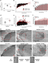
IFB-088 treatment improves MpzR98C/ + mice quadriceps femoral nerve morphology. a g-ratios performed on toluidine blue–stained semi-thin sections of quadriceps femoral nerve from MpzR98C/ + mice treated with vehicle b.i.d. or IFB-088 1 mg/kg b.i.d. for 5 months. Data are expressed as mean ± SEM. n = 4 mice per condition. *P < 0.05 by Student’s t-test. b Scatter plot of quadriceps femoral nerve g-ratios toward the axon diameter from MpzR98C/ + mice treated with vehicle b.i.d. or IFB-088 1 mg/kg b.i.d. n = 4 mice per condition. c Average g-ratio plotted by axon diameter. *P < 0.05 by Student’s t-test. d Myelin thickness measured from toluidine blue–stained semi-thin sections of quadriceps femoral nerve from MpzR98C/ + mice treated with vehicle b.i.d. or IFB-088 1 mg/kg b.i.d. for 5 months. Data are expressed in µm as mean ± SEM. n = 4 mice per condition. *P < 0.05 by Student’s t-test. e Scatter plot of quadriceps femoral nerve myelin thickness toward axon diameter from MpzR98C/ + mice treated with vehicle b.i.d. or IFB-088 1 mg/kg b.i.d. n = 4 mice per condition. f Average myelin thickness plotted by axon diameter. *P < 0.05 by Student’s t-test. g Ultrathin electron microscopy sections of quadriceps femoral nerve from WT mice treated with vehicle b.i.d. and from MpzR98C/ + mice treated with vehicle b.i.d. or IFB-088 1 mg/kg b.i.d. for 5 months. Lower panels are higher magnification images of upper panels. Scale bar 1 µm for top panels, and 500 nm for bottom panels. The insets show normal myelin compaction and periodicity
Fig. 3
Fig. 3
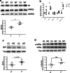
IFB-088 treatment reduces ER stress and Schwann cell differentiation marker expression in MpzR98C/ + sciatic nerve. Evaluation of mRNA (a–c) and protein levels (d–f) on sciatic nerve samples from WT and MpzR98C/ + mice treated with vehicle b.i.d. or IFB-088 1 mg/kg b.i.d. for 5 months. mRNA relative levels of Bip (a), Chop (b) and Xbp1s (c) determined by qRT-PCR. n = 4 per condition. #P < 0.05 by Mann–Whitney. d Western blot images for BiP, P-eIF2α, and c-Jun. Quantification relative to tubulin for P-IeF2α (e), BiP (f) and c-Jun (g), n = 3–5 per condition. *P < 0.05, **P < 0.01 by Student’s t-test
Fig. 4
Fig. 4
C3-PMP22 mice show a relative overexpression of PMP22 associated with the expression of ER stress/UPR markers. a Evaluation of PMP22, P0 and GAPDH protein levels by WB in sciatic nerve protein lysates from 4-month-old WT and C3-PMP22 (C3) mice. Top: representative picture. Bottom: quantification of the PMP22/P0 protein ratio; n = 3 per condition. *P < 0.05 by Student’s t-test. b Evaluation of Bip, Chop, Xbp1s, Atf4 and Gadd34/Ppp1r15a mRNA levels by qRT-PCR on sciatic nerve samples from 4-month-old WT and C3-PMP22 (C3) mice; n = 4 per condition. #P < 0.05 Mann–Whitney. c Evaluation of BiP and tubulin protein levels by WB in sciatic nerve protein lysates from 4-month-old WT and C3-PMP22 (C3) mice. Top: Representative picture. Bottom: quantification relative to tubulin; n = 6 per condition. Student’s t-test. d Evaluation of P-eIF2α, total eIF2α and tubulin protein level by WB in sciatic nerve protein lysates from 1-month-old WT and C3-PMP22 (C3) mice. Top: representative picture. Bottom: quantification of the P-eiF2α level relative to total eIF2α; n = 6 per condition. **P < 0.01 by unpaired t-test
Fig. 5
Fig. 5
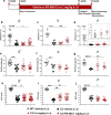
IFB-088 treatment improves motor function and nerve conduction velocity in C3-PMP22 mice. a Diagram of the treatment strategy. Fifteen-day-old WT and C3-PMP22 (C3) mice were orally administered with vehicle b.i.d. or IFB-088 at 0.5 or 1 mg/kg b.i.d. for 12 weeks. b Treadmill analysis performed after 10-week of treatment. Data from males and females expressed in meter (m) as mean ± SEM; n = 14–22 mice per condition. c Data from females; n = 8–10 mice per condition. d Data from males, n = 6–13 mice per condition. e Forepaws grip strength average of 6 trials performed after 10 weeks of treatment. Data were expressed in pull force (grams) as mean ± SEM. n = 14–22 mice per condition. f Data from females; n = 8–10 mice per condition. g Data from males, n = 6–13 mice per condition. h Motor nerve conduction velocity (MNCV) performed after 12 weeks of treatment. Data from males and females expressed in meters/second (m/s) as mean ± SEM. n = 14–21 mice per condition. (i) Data from females, n = 8–10 mice per condition. (j) Data from males, n = 6–12 mice per condition. *P < 0.05; **P < 0.01; ****P < 0.0001 by Student’s t-test. #P < 0.05; ###P < 0.001; ####P < 0.0001 by Mann–Whitney. &P < 0.05; &&P < 0.01 by one-way ANOVA followed by Dunnett’s test. $P < 0.05; $$P < 0.01; $$$P < 0.001 by Kruskal–Wallis followed by Dunn’s test
Fig. 6
Fig. 6
IFB-088 treatment improves C3-PMP22 mice quadriceps femoral nerve morphology. Toluidine blue–stained semithin sections of quadriceps femoral nerve from a female and b male WT mice treated with vehicle b.i.d. and C3-PMP22 (C3) mice treated with vehicle b.i.d. or IFB-088 at 0.5 or 1 mg/kg b.i.d. for 12 weeks. Scale bar, 10 µm; g-ratio measurement has been performed on toluidine blue–stained semi-thin sections. c Scatter plot of quadriceps femoral nerve g-ratios from WT mice treated with vehicle b.i.d. and C3-PMP22 (C3) mice treated with vehicle b.i.d. or IFB-088 at 0.5 or 1 mg/kg b.i.d. Note the “cloud” of axons with diameter lower than 1 µm present only in C3-PMP22 nerves (untreated and treated) and the considerable increase in the number of myelinated axons larger than 5–6 µm in IFB-088-treated C3-PMP22 nerves. n = 5–8 nerves per condition. d Percentage of myelinated axons per axons size; n = 5–8 nerves per condition. e, f TEM analysis of quadriceps femoral nerve from C3-PMP22 (C3) mice treated with vehicle b.i.d. or IFB-088 at 1 mg/kg b.i.d. e In C3-PMP22 vehicle–treated nerves, small-calibre axons are abnormally hypermyelinated (black dotted arrows). Treatment with IFB-088 results in normal-looking myelin in axons larger than 1 µm (red arrows) or in almost no myelin in smaller axons (blue larges arrows). f In C3-PMP22 vehicle–treated nerves, large-calibre axons are basically amyelinated or with only a very thin layer of myelin (left panel). After treatment with IFB-088 (right panel), a subgroup of large-calibre axons showed a properly compacted (albeit still rather thin) myelin sheath. A side-by-side comparison of myelination in two axons with similar calibre is shown in the lower panel
Fig. 7
Fig. 7
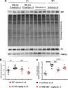
IFB-088 treatment improves PMP22 stoichiometry in C3-PMP22 mouse peripheral nerves and reduces ER stress. Evaluation of PMP22, P0 and BiP protein levels by WB in sciatic nerve protein lysates from WT mice treated with vehicle b.i.d. and C3-PMP22 (C3) mice treated with vehicle b.i.d. or IFB-088 at 0.5 or 1 mg/kg b.i.d. a Representative picture. Three representative samples per condition out of five/six are shown. b Quantification of BiP protein level. c Quantification of the PMP22/P0 protein ratio. *P < 0.05; **P < 0.01; ****P < 0.0001 by Student’s t-test

Similar articles

Cited by

References

    1. Skre H. Genetic and clinical aspects of Charcot-Marie-Tooth's disease. Clin Genet. 1974;6:98–118. doi: 10.1111/j.1399-0004.1974.tb00638.x. - DOI - PubMed
    1. Fridman V, Bundy B, Reilly MM, Pareyson D, Bacon C, Burns J, Day J, Feely S, et al. CMT subtypes and disease burden in patients enrolled in the Inherited Neuropathies Consortium natural history study: a cross-sectional analysis. J Neurol Neurosurg Psychiatry. 2015;86:873–878. doi: 10.1136/jnnp-2014-308826. - DOI - PMC - PubMed
    1. Murphy SM, Laura M, Fawcett K, Pandraud A, Liu YT, Davidson GL, Rossor AM, Polke JM, et al. Charcot-Marie-Tooth disease: frequency of genetic subtypes and guidelines for genetic testing. J Neurol Neurosurg Psychiatry. 2012;83:706–710. doi: 10.1136/jnnp-2012-302451. - DOI - PMC - PubMed
    1. Lupski JR, de Oca-Luna RM, Slaugenhaupt S, Pentao L, Guzzetta V, Trask BJ, Saucedo-Cardenas O, Barker DF, et al. DNA duplication associated with Charcot-Marie-Tooth disease type 1A. Cell. 1991;66:219–232. doi: 10.1016/0092-8674(91)90613-4. - DOI - PubMed
    1. Timmerman V, Nelis E, Van Hul W, Nieuwenhuijsen BW, Chen KL, Wang S, Ben Othman K, Cullen B, et al. The peripheral myelin protein gene PMP-22 is contained within the Charcot-Marie-Tooth disease type 1A duplication. Nat Genet. 1992;1:171–175. doi: 10.1038/ng0692-171. - DOI - PubMed

MeSH terms

Substances