

Clinically undetected plasmacytoid urothelial carcinoma of the urinary bladder with non-mass-forming metastases in multiple organs: an autopsy case
- PMID: 35501670
- PMCID: PMC9288892
- DOI: 10.4132/jptm.2022.03.15
Clinically undetected plasmacytoid urothelial carcinoma of the urinary bladder with non-mass-forming metastases in multiple organs: an autopsy case
Abstract
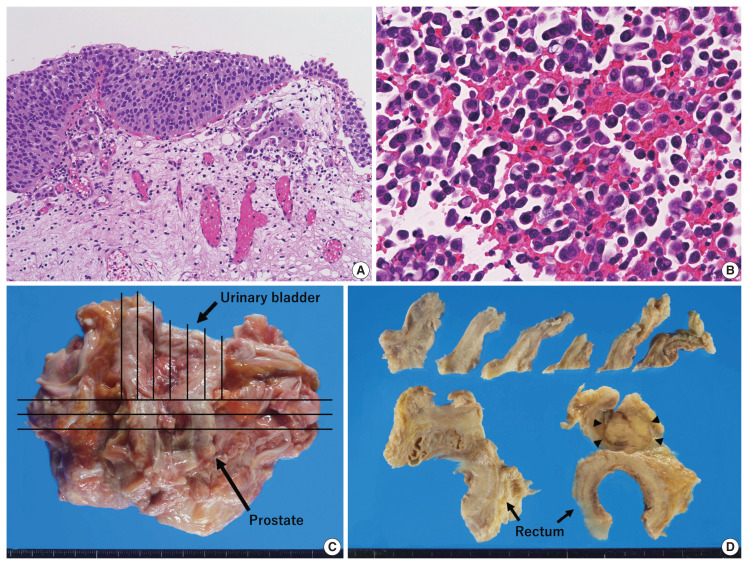
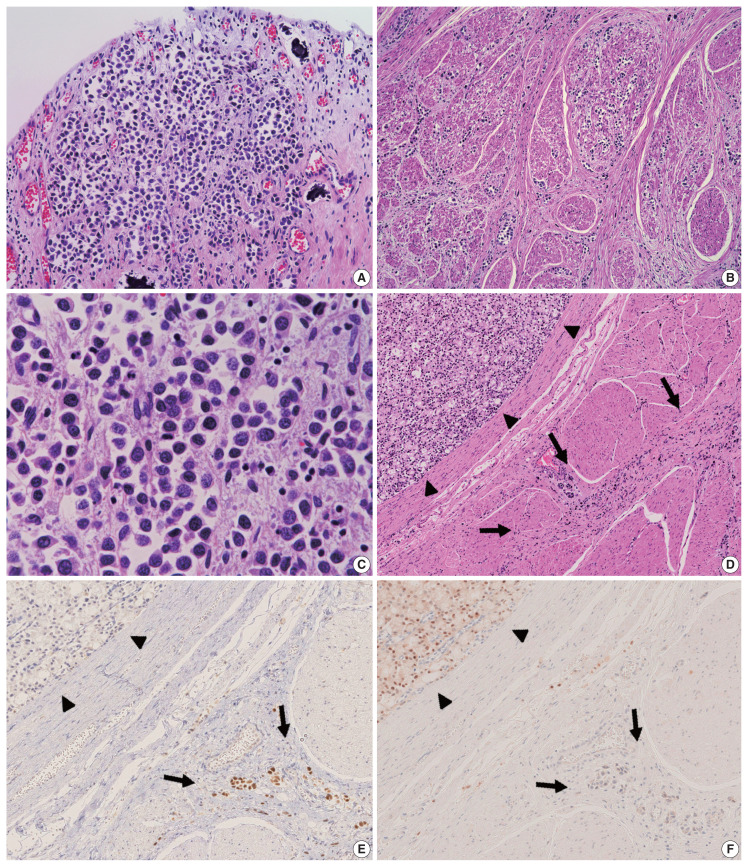
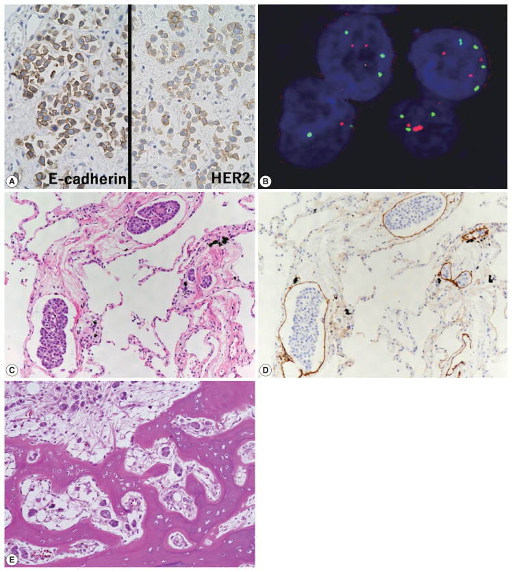
This case report outlines a clinically undetected urinary bladder plasmacytoid urothelial carcinoma (PUC) with multiple metastases detected at autopsy. An 89-year-old man presented with edema in the lower limbs. Pleural fluid cytology revealed discohesive carcinomatous cells, although imaging studies failed to identify the primary site of tumor. The patient died of respiratory failure. Autopsy disclosed a prostate tumor and diffusely thickened urinary bladder and rectum without distinct tumorous lesions. Histologically, the tumor consisted of acinar-type prostate adenocarcinoma with no signs of metastasis. Additionally, small, plasmacytoid tumor cells were observed in the urinary bladder/rectum as isolated or small clustering fashions. These metastasized to the lungs, intestine, generalized lymph nodes in a non-mass-forming manner. Combined with immunohistochemical studies, these tumor cells were diagnosed PUC derived from the urinary bladder. Both clinicians and pathologists should recognize PUC as an aggressive histological variant, which can represent a rapid systemic progression without mass-forming lesions.
Keywords: Autopsy; Cancer of unknown primary; Plasmacytoid variant; Urinary bladder; Urothelial carcinoma.
Conflict of interest statement
The authors declare that they have no potential conflicts of interest.
Figures
Similar articles
-
Carcinomatous Meningitis and Hydrocephalus in Plasmacytoid Urothelial Carcinoma of the Urinary Bladder With Extremely Elevated CA19-9 Levels.Cureus. 2024 Feb 21;16(2):e54643. doi: 10.7759/cureus.54643. eCollection 2024 Feb. Cureus. 2024. PMID: 38523920 Free PMC article.
-
Plasmacytoid urothelial carcinoma of the urinary bladder report of seven new cases.Eur Urol. 2006 Nov;50(5):1111-4. doi: 10.1016/j.eururo.2005.12.047. Epub 2006 Jan 18. Eur Urol. 2006. PMID: 16626859
-
Plasmacytoid urothelial carcinoma of the urinary bladder: a clinical pathological study and literature review.Int J Clin Exp Pathol. 2012;5(6):601-8. Epub 2012 Jul 29. Int J Clin Exp Pathol. 2012. PMID: 22949945 Free PMC article.
-
[Plasmacytoid urothelial carcinoma of the urinary bladder. A study of 7 cases].Actas Urol Esp. 2008 Sep;32(8):806-10. doi: 10.1016/s0210-4806(08)73939-1. Actas Urol Esp. 2008. PMID: 19013979 Review. Spanish.
-
Plasmacytoid Urothelial Carcinoma of Ureter with Retroperitoneal Metastasis: A Case Report.Am J Case Rep. 2018 Feb 13;19:158-162. doi: 10.12659/ajcr.906679. Am J Case Rep. 2018. PMID: 29434183 Free PMC article. Review.
Cited by
-
Carcinomatous Meningitis and Hydrocephalus in Plasmacytoid Urothelial Carcinoma of the Urinary Bladder With Extremely Elevated CA19-9 Levels.Cureus. 2024 Feb 21;16(2):e54643. doi: 10.7759/cureus.54643. eCollection 2024 Feb. Cureus. 2024. PMID: 38523920 Free PMC article.
-
Severe Rectal Stenosis as the First Clinical Appearance of a Metastasis Originating from the Bladder: A Case Report and Literature Review.Life (Basel). 2025 Apr 22;15(5):682. doi: 10.3390/life15050682. Life (Basel). 2025. PMID: 40430111 Free PMC article. Review.
-
Plasmacytoid urothelial carcinoma: a multidisciplinary approach to the diagnosis and management.Abdom Radiol (NY). 2024 Sep 17. doi: 10.1007/s00261-024-04553-9. Online ahead of print. Abdom Radiol (NY). 2024. PMID: 39287629 Review. No abstract available.
References
-
- Sahin AA, Myhre M, Ro JY, Sneige N, Dekmezian RH, Ayala AG. Plasmacytoid transitional cell carcinoma: report of a case with initial presentation mimicking multiple myeloma. Acta Cytol. 1991;35:277–80. - PubMed
-
- Zukerberg LR, Harris NL, Young RH. Carcinomas of the urinary bladder simulating malignant lymphoma: a report of five cases. Am J Surg Pathol. 1991;15:569–76. - PubMed
-
- Ro JY, Shen SS, Lee HI, et al. Plasmacytoid transitional cell carcinoma of urinary bladder: a clinicopathologic study of 9 cases. Am J Surg Pathol. 2008;32:752–7. - PubMed
-
- Kim DK, Kim JW, Ro JY, et al. Plasmacytoid variant urothelial carcinoma of the bladder: a systematic review and meta-analysis of clinicopathological features and survival outcomes. J Urol. 2020;204:215–23. - PubMed
-
- Sood S, Paner GP. Plasmacytoid urothelial carcinoma: an unusual variant that warrants aggressive management and critical distinction on transurethral resections. Arch Pathol Lab Med. 2019;143:1562–7. - PubMed
LinkOut - more resources
Full Text Sources
