. 2022 Apr 30;22(1):80.
doi: 10.1186/s12880-022-00808-3.

The efficacy of deep learning models in the diagnosis of endometrial cancer using MRI: a comparison with radiologists

Affiliations

The efficacy of deep learning models in the diagnosis of endometrial cancer using MRI: a comparison with radiologists

Aiko Urushibara et al. BMC Med Imaging. .

Abstract

Purpose: To compare the diagnostic performance of deep learning models using convolutional neural networks (CNN) with that of radiologists in diagnosing endometrial cancer and to verify suitable imaging conditions.

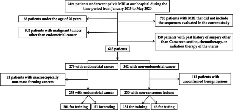
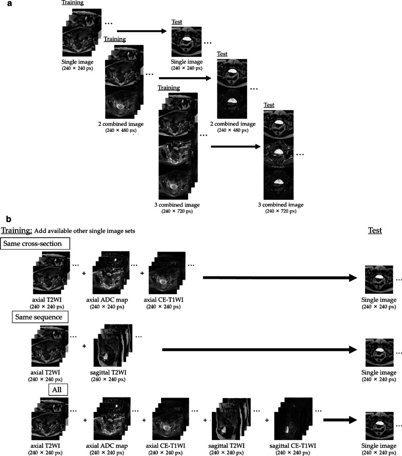
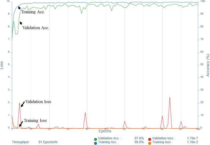
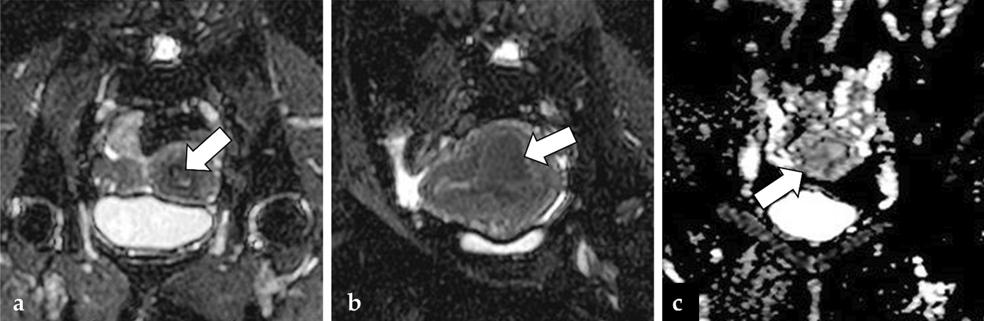
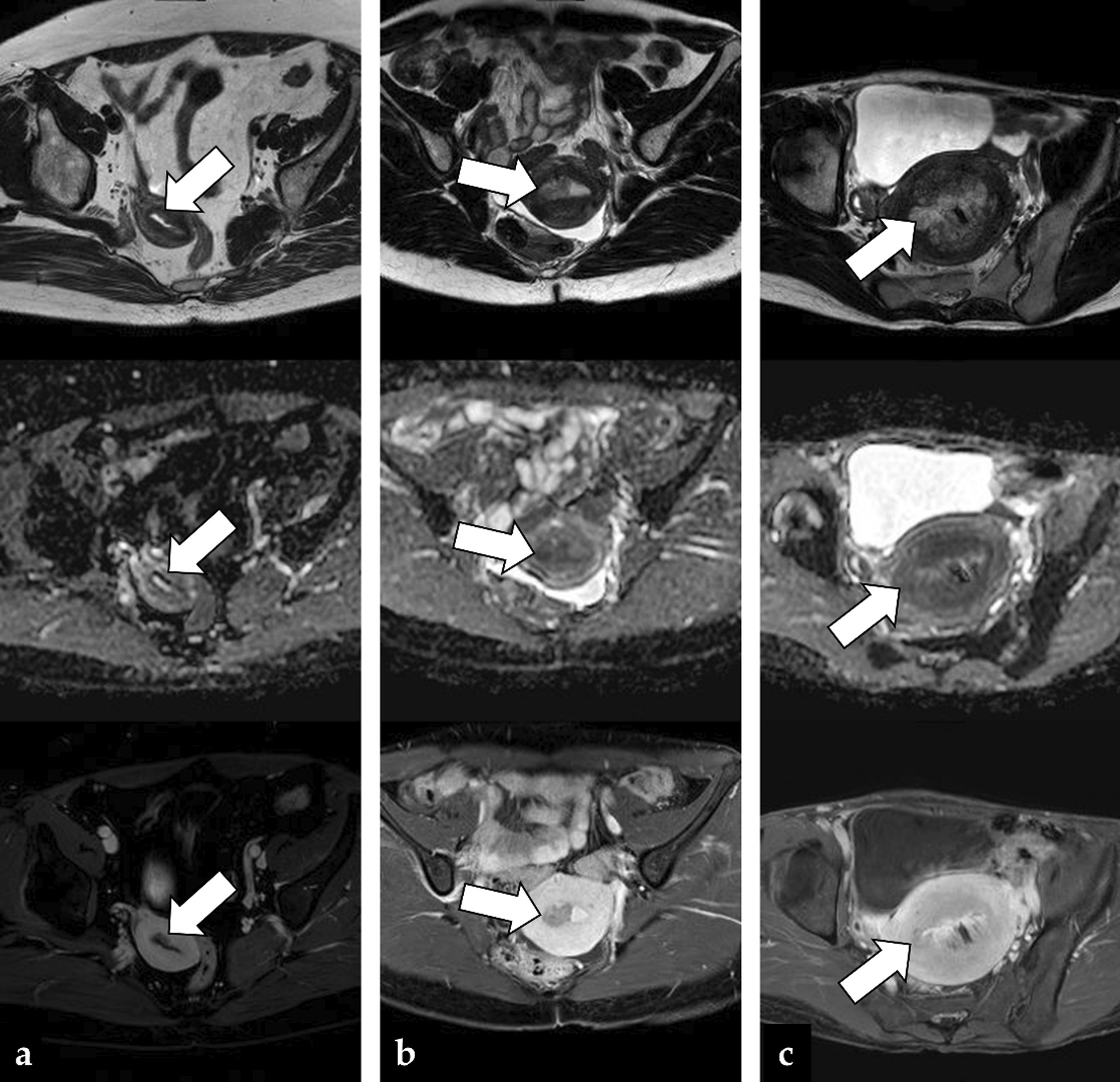
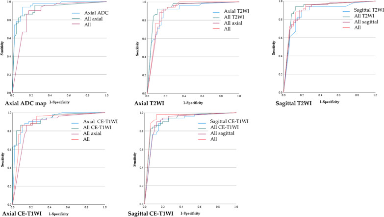
Methods: This retrospective study included patients with endometrial cancer or non-cancerous lesions who underwent MRI between 2015 and 2020. In Experiment 1, single and combined image sets of several sequences from 204 patients with cancer and 184 patients with non-cancerous lesions were used to train CNNs. Subsequently, testing was performed using 97 images from 51 patients with cancer and 46 patients with non-cancerous lesions. The test image sets were independently interpreted by three blinded radiologists. Experiment 2 investigated whether the addition of different types of images for training using the single image sets improved the diagnostic performance of CNNs.

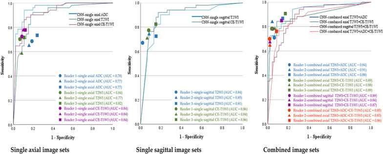
Results: The AUC of the CNNs pertaining to the single and combined image sets were 0.88-0.95 and 0.87-0.93, respectively, indicating non-inferior diagnostic performance than the radiologists. The AUC of the CNNs trained with the addition of other types of single images to the single image sets was 0.88-0.95.

Conclusion: CNNs demonstrated high diagnostic performance for the diagnosis of endometrial cancer using MRI. Although there were no significant differences, adding other types of images improved the diagnostic performance for some single image sets.

Keywords: Artificial intelligence; CNN; Convolutional neural network; Endometrial carcinoma; Magnetic resonance imaging.


Conflict of interest statement

The authors declare that they have no competing interests.

Figures

Fig. 1
Fig. 1
Flowchart of the patient selection process
Fig. 2
Fig. 2
a Schematic diagrams of Experiment 1. b Schematic diagrams of Experiment 2. T2WI, T2 weighted image; ADC, Apparent Diffusion Coefficient; CE-T1WI, contrast-enhanced T1 weighted image
Fig. 3
Fig. 3
Experiment 1-The ROC curves for the CNNs. The ROC curves for the CNNs pertaining to the testing of the single and combined image sets with the AUC plots for the radiologists. T2WI, T2 weighted image; ADC, Apparent Diffusion Coefficient; CE-T1WI, contrast-enhanced T1 weighted image
Fig. 4
Fig. 4
Accuracy and loss of the training data of the single image set of axial ADC map. Accuracy and loss of the training data of the single image set of axial ADC map with the training/validation split ratio 9:1 and epoch 100 in Experiment 1. Acc., accuracy
Fig. 5
Fig. 5
Three cases of false negatives were observed in the single image set of axial ADC: a A 55-year-old woman with grade 1 endometrioid carcinoma, in which the CNN was able to diagnose cancer, but the readers 1, 2, and 3 were not (the CNN confidence; cancer = 99.9%). The image shows a tiny tumor filling the uterine cavity (arrow); b A 34-year-old woman with grade 1 endometrioid carcinoma, in which all the three readers could diagnose cancer, but the CNN could not (the CNN confidence; cancer = 18.8%). The image shows a massive tumor protruding into the myometrium of the posterior wall of the uterus (arrow); c A 31-year-old woman with grade 2 endometrioid carcinoma, in which neither the CNN nor the three readers could diagnose the presence of cancer (the CNN confidence; cancer = 22.5%). The image shows the tumor filling the uterine cavity (arrow). A slight decrease in the single image of ADC map might have made the diagnosis of tumor difficult with a single image without considering the other images for radiologists
Fig. 6
Fig. 6
Three cases of false negatives were observed in the combined image set of axial T2WI + ADC + CE-T1WI: a A 56-year-old woman with grade 1 endometrioid carcinoma, in which the CNN was able to detect the cancer, but the three readers were not (the CNN confidence; cancer = 100%); b A 30-year-old woman with grade 1 endometrioid carcinoma, in which the three readers could diagnose the presence of cancer, but the CNN could not (the CNN confidence; cancer = 0.5%). The image shows a tumor displaying the typical appearance of endometrial cancer and filling the right side of the uterine cavity (arrow); c A 45-year-old woman with grade 1 endometrioid carcinoma, in which neither the CNN nor the three readers could diagnose the presence of cancer (the CNN confidence; cancer = 0.5%). The image shows a massive tumor filling the uterine cavity (arrow) and a hemorrhage at the center of the lesion. Non-uniform signal intensities of the tumor mass may have made the diagnosis difficult for radiologists
Fig. 7
Fig. 7
Experiment 2—The ROC curves for the CNNs. The ROC curves for the CNNs pertaining to testing the single image sets with various types of image sets for training. ADC, Apparent Diffusion Coefficient; T2WI, T2 weighted image; CE-T1WI, contrast-enhanced T1 weighted image

Similar articles

Cited by

References

    1. Sung H, Ferlay J, Siegel RL, Laversanne M, Soerjomataram I, Jemal A, et al. Global Cancer Statistics 2020: GLOBOCAN estimates of incidence and mortality worldwide for 36 cancers in 185 countries. CA Cancer J Clin. 2021;71(3):209–249. doi: 10.3322/caac.21660. - DOI - PubMed
    1. Constantine GD, Kessler G, Graham S, Goldstein SR. Increased incidence of endometrial cancer following the women's health initiative: an assessment of risk factors. J Womens Health (Larchmt) 2019;28(2):237–243. doi: 10.1089/jwh.2018.6956. - DOI - PMC - PubMed
    1. Sala E, Wakely S, Senior E, Lomas D. MRI of malignant neoplasms of the uterine corpus and cervix. AJR Am J Roentgenol. 2007;188(6):1577–1587. doi: 10.2214/AJR.06.1196. - DOI - PubMed
    1. Beddy P, Moyle P, Kataoka M, Yamamoto AK, Joubert I, Lomas D, et al. Evaluation of depth of myometrial invasion and overall staging in endometrial cancer: comparison of diffusion-weighted and dynamic contrast-enhanced MR imaging. Radiology. 2012;262(2):530–537. doi: 10.1148/radiol.11110984. - DOI - PubMed
    1. Nougaret S, Horta M, Sala E, Lakhman Y, Thomassin-Naggara I, Kido A, et al. Endometrial cancer MRI staging: updated guidelines of the European society of urogenital radiology. Eur Radiol. 2019;29(2):792–805. doi: 10.1007/s00330-018-5515-y. - DOI - PubMed

LinkOut - more resources