. 2022 May 2;22(1):198.
doi: 10.1186/s12886-022-02420-z.

Vascular tortuosity analysis in eyes with epiretinal membrane imaged by optical coherence tomography angiography

Affiliations

Vascular tortuosity analysis in eyes with epiretinal membrane imaged by optical coherence tomography angiography

Kosuke Miyazawa et al. BMC Ophthalmol. .

Abstract

Background: This study aimed to evaluate macular vessel tortuosity using optical coherence tomography angiography (OCTA) and its association with visual outcomes in eyes undergoing surgery for epiretinal membrane (ERM).

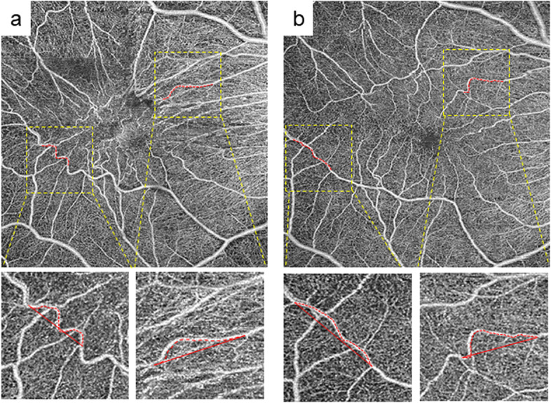
Methods: The study included 22 consecutive patients who underwent vitrectomy for ERM between May 2019 and July 2020 and OCTA at Osaka University Hospital. All patients underwent ophthalmologic examinations, including swept-source OCTA. Standard vitrectomy was performed, and the patients were followed up for 6 months postoperatively. Distortion of retinal vessels was calculated using two parameters: the actual vessel length in the vessel section (VL) and the direct vessel branching point distance (BD) in the three quadrants (nasal, temporal, and superior-inferior) of the macula. We analyzed the correlation between these parameters and visual outcomes.

Results: Significantly longer VL was found at 1, 3, and 6 months postoperatively (p = 0.006, 0.008, and 0.022, respectively) in the temporal quadrant compared to baseline temporal VL. Significantly shorter VL was found in nasal quadrants at 1 and 3 months (p = 0.046 and p = 0.018) in the comparison of nasal baseline VL. VL/BDs were correlated with the same postoperative best-corrected visual acuity (BCVA) at 1, 3, and 6 months (p = 0.035, 0.035, and 0.042, respectively) in the superior-inferior quadrant. A significant association of changes in VL and BCVA was found at 3 and 6 months postoperatively in the nasal quadrant (p = 0.018 and 0.0455, respectively).

Conclusions: Changes in vascular distortion after ERM surgery can be measured using OCTA. The change in vessels around the macula became more linear; this was associated with visual outcomes after surgery.

Keywords: Epiretinal membrane; Imaging analysis; Optical coherence tomography angiography; Vascular tortuosity; Vitrectomy.


Conflict of interest statement

The authors declare that they have no competing interests.

Figures

Fig. 1
Fig. 1
Representative en face image of optical coherence tomography angiography (OCTA) shows microvascular structure around the macula in a right eye with epiretinal membrane. a A 6 × 6 mm en face image of OCTA is divided into four quadrants. b A high magnification image of the red square in (a). A retinal vessel was randomly picked in each quadrant to macula descending to macula. We also picked two adjacent branch points. Red dotted line represents the actual vessel length in the vessel section and yellow line represents the direct vessel branching point distance between two adjacent branch points
Fig. 2
Fig. 2
Representative en face images of preoperative and postoperative optical coherence tomography angiography (OCTA) in eyes with ERM. a Preoperative and b postoperative 1 month 6 × 6 mm OCTA images. Red dotted lines show temporal and nasal vessels analyzed in this study. High magnification images are shown below, which correspond to the dotted yellow square. The length of red dotted lines defined as “the actual vessel length (VL)” and the length of red lines defined as “the direct vessel branching point distance (BD),” respectively. Note that VL becomes shorter in temporal vessels and longer in nasal vessels postoperatively. In contrast, BD becomes longer in temporal and shorter in nasal postoperatively
Fig. 3
Fig. 3
Scatter plots of data summary described in Tables 3 and 4. a Scatter plots depicting the association between superior/inferior VL/BD and BCVA postoperatively at 1 month (1 M), 3 months (3 M), and 6 months (6 M), which was shown in Table 3. Lines show regression lines. b Scatter plots depicting the association between the change of VL in the nasal quadrant and the change of BCVA postoperatively at 1 month (1 M), 3 months (3 M), and 6 months (6 M), which was shown in Table 4. Lines show regression lines. VL: the actual vessel length, BD: the direct vessel branching point distance, BCVA: best-corrected visual acuity

Similar articles

Cited by

References

    1. Kampik A, Green WR, Michels RG, Nase PK. Ultrastructural features of progressive idiopathic epiretinal membrane removed by vitreous surgery. Am J Ophthalmol. 1980;90:797–809. doi: 10.1016/S0002-9394(14)75195-5. - DOI - PubMed
    1. Wilkins JR, Puliafito CA, Hee MR, Duker JS, Reichel E, Coker JG, et al. Characterization of epiretinal membranes using optical coherence tomography. 1996;103:2142–2151. - PubMed
    1. Grewing R, Mester U. Results of surgery for epiretinal membranes and their recurrences. Br J Ophthalmol. 1996;80:323–326. doi: 10.1136/bjo.80.4.323. - DOI - PMC - PubMed
    1. Massin P, Allouch C, Haouchine B, Metge F, Paques M, Tangui L, et al. Optical coherence tomography of idiopathic macular epiretinal membranes before and after surgery. Am J Ophthalmol. 2000;130:732–739. doi: 10.1016/S0002-9394(00)00574-2. - DOI - PubMed
    1. Govetto A, Lalane RA, 3rd, Sarraf D, Figueroa MS, Hubschman JP. Insights into epiretinal membranes: presence of ectopic inner foveal layers and a new optical coherence tomography staging scheme. Am J Ophthalmol. 2017;175:99–113. doi: 10.1016/j.ajo.2016.12.006. - DOI - PubMed

LinkOut - more resources