

Central and peripheral contributions of T-type calcium channels in pain
- PMID: 35501819
- PMCID: PMC9063214
- DOI: 10.1186/s13041-022-00923-w
Central and peripheral contributions of T-type calcium channels in pain
Abstract
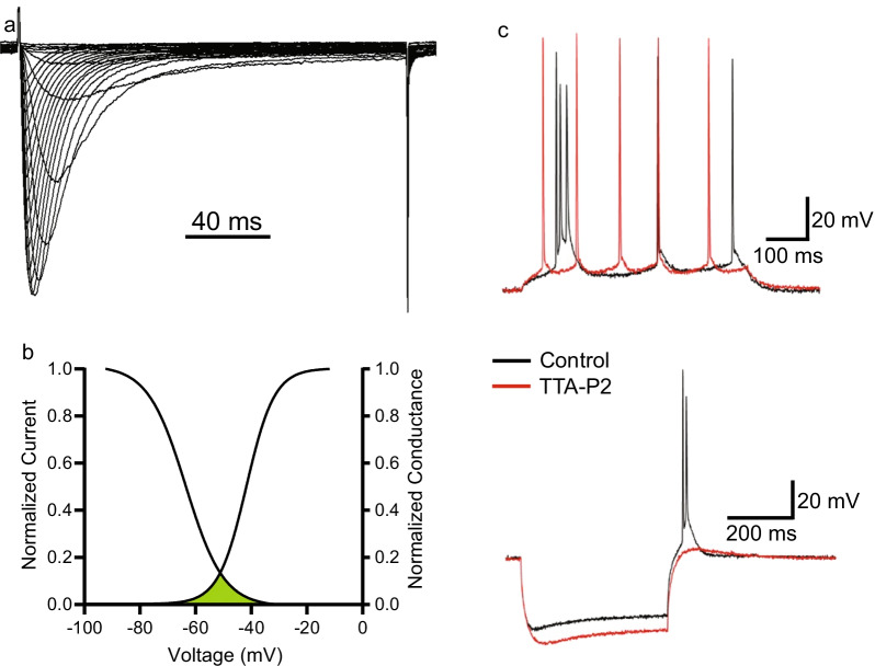
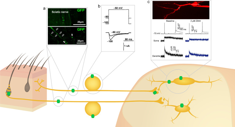
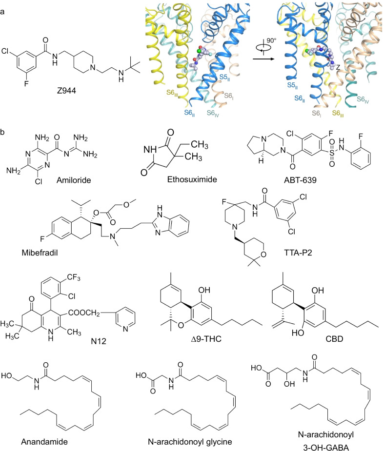
Chronic pain is a severely debilitating condition that reflects a long-term sensitization of signal transduction in the afferent pain pathway. Among the key players in this pathway are T-type calcium channels, in particular the Cav3.2 isoform. Because of their biophysical characteristics, these channels are ideally suited towards regulating neuronal excitability. Recent evidence suggests that T-type channels contribute to excitability of neurons all along the ascending and descending pain pathways, within primary afferent neurons, spinal dorsal horn neurons, and within pain-processing neurons in the midbrain and cortex. Here we review the contribution of T-type channels to neuronal excitability and function in each of these neuronal populations and how they are dysregulated in chronic pain conditions. Finally, we discuss their molecular pharmacology and the potential role of these channels as therapeutic targets for chronic pain.
Keywords: Analgesia; CACNA1H; Cav3.2; Glycosylation; Pain; T-type; Ubiquitination.
© 2022. The Author(s).
Conflict of interest statement
The authors declare no competing interest.
Figures
Similar articles
-
Orai1 Plays a Crucial Role in Central Sensitization by Modulating Neuronal Excitability.J Neurosci. 2018 Jan 24;38(4):887-900. doi: 10.1523/JNEUROSCI.3007-17.2017. Epub 2017 Dec 11. J Neurosci. 2018. PMID: 29229703 Free PMC article.
-
Targeting T-type/CaV3.2 channels for chronic pain.Transl Res. 2021 Aug;234:20-30. doi: 10.1016/j.trsl.2021.01.002. Epub 2021 Jan 7. Transl Res. 2021. PMID: 33422652 Free PMC article. Review.
-
Cdk5-Dependent Phosphorylation of CaV3.2 T-Type Channels: Possible Role in Nerve Ligation-Induced Neuropathic Allodynia and the Compound Action Potential in Primary Afferent C Fibers.J Neurosci. 2020 Jan 8;40(2):283-296. doi: 10.1523/JNEUROSCI.0181-19.2019. Epub 2019 Nov 19. J Neurosci. 2020. PMID: 31744861 Free PMC article.
-
Inflammation reduces the contribution of N-type calcium channels to primary afferent synaptic transmission onto NK1 receptor-positive lamina I neurons in the rat dorsal horn.J Physiol. 2007 May 1;580(Pt.3):883-94. doi: 10.1113/jphysiol.2006.125880. Epub 2007 Feb 15. J Physiol. 2007. PMID: 17303639 Free PMC article.
-
T-type calcium channels: functional regulation and implication in pain signaling.J Pharmacol Sci. 2013;122(4):244-50. doi: 10.1254/jphs.13r05cp. Epub 2013 Aug 1. J Pharmacol Sci. 2013. PMID: 23903007 Review.
Cited by
-
Betulinic acid analogs inhibit N- and T-type voltage-gated calcium channels to attenuate nerve-injury associated neuropathic and formalin models of pain.Neurobiol Pain. 2023 Jan 14;13:100116. doi: 10.1016/j.ynpai.2023.100116. eCollection 2023 Jan-Jul. Neurobiol Pain. 2023. PMID: 36687466 Free PMC article.
-
RNA 5-Methylcytosine regulators are associated with cell adhesion and predict prognosis of endometrial cancer.Transl Cancer Res. 2023 Oct 31;12(10):2556-2571. doi: 10.21037/tcr-23-742. Epub 2023 Oct 24. Transl Cancer Res. 2023. PMID: 37969377 Free PMC article.
-
Electrophysiological characterization of a CaV3.1 calcium channel mutation linked to trigeminal neuralgia.Pflugers Arch. 2023 Jun;475(6):711-718. doi: 10.1007/s00424-023-02808-w. Epub 2023 Apr 3. Pflugers Arch. 2023. PMID: 37010626
-
The anticonvulsant phytocannabinoids CBGVA and CBDVA inhibit recombinant T-type channels.Front Pharmacol. 2022 Nov 1;13:1048259. doi: 10.3389/fphar.2022.1048259. eCollection 2022. Front Pharmacol. 2022. PMID: 36386164 Free PMC article.
-
Pharmacogenetic landscape of pain management variants among Mediterranean populations.Front Pharmacol. 2024 May 15;15:1380613. doi: 10.3389/fphar.2024.1380613. eCollection 2024. Front Pharmacol. 2024. PMID: 38813106 Free PMC article.
References
Publication types
MeSH terms
Substances
Grants and funding
LinkOut - more resources
Full Text Sources
Other Literature Sources
Medical
