

An RNA-sequencing transcriptome of the rodent Schwann cell response to peripheral nerve injury
- PMID: 35501870
- PMCID: PMC9063194
- DOI: 10.1186/s12974-022-02462-6
An RNA-sequencing transcriptome of the rodent Schwann cell response to peripheral nerve injury
Abstract
Background: The important contribution of glia to mechanisms of injury and repair of the nervous system is increasingly recognized. In stark contrast to the central nervous system (CNS), the peripheral nervous system (PNS) has a remarkable capacity for regeneration after injury. Schwann cells are recognized as key contributors to PNS regeneration, but the molecular underpinnings of the Schwann cell response to injury and how they interact with the inflammatory response remain incompletely understood.
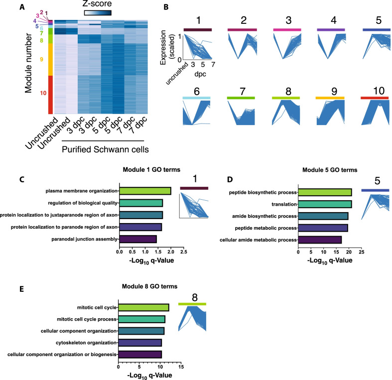
Methods: We completed bulk RNA-sequencing of Schwann cells purified acutely using immunopanning from the naïve and injured rodent sciatic nerve at 3, 5, and 7 days post-injury. We used qRT-PCR and in situ hybridization to assess cell purity and probe dataset integrity. Finally, we used bioinformatic analysis to probe Schwann cell-specific injury-induced modulation of cellular pathways.
Results: Our data confirm Schwann cell purity and validate RNAseq dataset integrity. Bioinformatic analysis identifies discrete modules of genes that follow distinct patterns of regulation in the 1st days after injury and their corresponding molecular pathways. These findings enable improved differentiation of myeloid and glial components of neuroinflammation after peripheral nerve injury and highlight novel molecular aspects of the Schwann cell injury response such as acute downregulation of the AGE/RAGE pathway and of secreted molecules Sparcl1 and Sema5a.
Conclusions: We provide a helpful resource for further deciphering the Schwann cell injury response and a depth of transcriptional data that can complement the findings of recent single cell sequencing approaches. As more data become available on the response of CNS glia to injury, we anticipate that this dataset will provide a valuable platform for understanding key differences in the PNS and CNS glial responses to injury and for designing approaches to ameliorate CNS regeneration.
Keywords: Injury response; Macrophage; Neuroinflammation; Peripheral nerve; Regeneration; Repair cell; Schwann cell; Transcriptome.
© 2022. The Author(s).
Conflict of interest statement
The authors declare no conflicts of interests.
Figures


Similar articles
-
The Glia Response after Peripheral Nerve Injury: A Comparison between Schwann Cells and Olfactory Ensheathing Cells and Their Uses for Neural Regenerative Therapies.Int J Mol Sci. 2017 Jan 29;18(2):287. doi: 10.3390/ijms18020287. Int J Mol Sci. 2017. PMID: 28146061 Free PMC article. Review.
-
Schwann cells use TAM receptor-mediated phagocytosis in addition to autophagy to clear myelin in a mouse model of nerve injury.Proc Natl Acad Sci U S A. 2017 Sep 19;114(38):E8072-E8080. doi: 10.1073/pnas.1710566114. Epub 2017 Sep 5. Proc Natl Acad Sci U S A. 2017. PMID: 28874532 Free PMC article.
-
A Schwann cell-enriched circular RNA circ-Ankib1 regulates Schwann cell proliferation following peripheral nerve injury.FASEB J. 2019 Nov;33(11):12409-12424. doi: 10.1096/fj.201900965R. Epub 2019 Sep 16. FASEB J. 2019. PMID: 31415184 Free PMC article.
-
Wallerian degeneration: gaining perspective on inflammatory events after peripheral nerve injury.J Neuroinflammation. 2011 Aug 30;8:110. doi: 10.1186/1742-2094-8-110. J Neuroinflammation. 2011. PMID: 21878126 Free PMC article. Review.
-
Down-regulation miR-146a-5p in Schwann cell-derived exosomes induced macrophage M1 polarization by impairing the inhibition on TRAF6/NF-κB pathway after peripheral nerve injury.Exp Neurol. 2023 Apr;362:114295. doi: 10.1016/j.expneurol.2022.114295. Epub 2022 Dec 6. Exp Neurol. 2023. PMID: 36493861
Cited by
-
The CXCL12-CXCR4-NLRP3 axis promotes Schwann cell pyroptosis and sciatic nerve demyelination in rats.Clin Exp Immunol. 2023 Dec 12;214(2):219-234. doi: 10.1093/cei/uxad081. Clin Exp Immunol. 2023. PMID: 37497691 Free PMC article.
-
Effects of PEMF and LIPUS Therapy on the Expression of Genes Related to Peripheral Nerve Regeneration in Schwann Cells.Int J Mol Sci. 2024 Nov 28;25(23):12791. doi: 10.3390/ijms252312791. Int J Mol Sci. 2024. PMID: 39684499 Free PMC article.
-
MNK1 and MNK2 expression in the human dorsal root and trigeminal ganglion.bioRxiv [Preprint]. 2023 Jan 4:2023.01.04.522773. doi: 10.1101/2023.01.04.522773. bioRxiv. 2023. Update in: Neuroscience. 2023 Apr 1;515:96-107. doi: 10.1016/j.neuroscience.2023.01.039. PMID: 36711529 Free PMC article. Updated. Preprint.
-
EZH2-dependent myelination following sciatic nerve injury.Neural Regen Res. 2025 Aug 1;20(8):2382-2394. doi: 10.4103/NRR.NRR-D-23-02040. Epub 2024 May 13. Neural Regen Res. 2025. PMID: 39359095 Free PMC article.
-
Unveiling the Mechanisms of Pain in Endometriosis: Comprehensive Analysis of Inflammatory Sensitization and Therapeutic Potential.Int J Mol Sci. 2025 Feb 19;26(4):1770. doi: 10.3390/ijms26041770. Int J Mol Sci. 2025. PMID: 40004233 Free PMC article. Review.
References
-
- Arthur-Farraj PJ, Morgan CC, Adamowicz M, Gomez-Sanchez JA, Fazal SV, Beucher A, et al. Changes in the coding and non-coding transcriptome and DNA methylome that define the schwann cell repair phenotype after nerve injury. Cell Rep. 2017;20(11):2719–2734. doi: 10.1016/j.celrep.2017.08.064. - DOI - PMC - PubMed
-
- Blighe K, Rana S, Lewis M. EnhancedVolcano: publication-ready volcano plots with enhanced colouring and labeling. R package version 1.10.0. 2021.
-
- Brosius Lutz A, Chung WS, Sloan SA, Carson GA, Zhou L, Lovelett E, et al. Schwann cells use TAM receptor-mediated phagocytosis in addition to autophagy to clear myelin in a mouse model of nerve injury. Proc Natl Acad Sci USA. 2017;114(38):E8072–e8080. doi: 10.1073/pnas.1710566114. - DOI - PMC - PubMed
MeSH terms
Substances
Grants and funding
LinkOut - more resources
Full Text Sources
Medical
Molecular Biology Databases
Research Materials
Miscellaneous
